They Survived the Holocaust to Battle Cancer

Holocaust memorial Boston

The Holocaust was the genocide of millions of Jews by the Nazis between the years 1939 and 1945. Researchers at Ben-Gurion University of the Negev in Israel published a study in 2018 that found a higher rate of cancer occurrence in Holocaust survivors in Israel compared to those who did not experience the Holocaust. There is very little information regarding cancer in this group of individuals. All legal decisions concerning financial assistance for Holocaust survivors fighting cancer is based only on one former study. Keeping this in mind, the researchers conducted a comprehensive study that took into account misleading cancer risk factors like age, obesity, diabetes, and smoking.

The study was conducted on 294,543 individuals who were Holocaust survivors based on recognition status as defined by the Holocaust Survivor Benefits Law. Data analysis was done on some of the most prevalent cancers in Israel, cancers of the lungs, colon, bladder, melanoma, breast, and prostate. Analysis for the last two cancers were conducted only on women and men respectively. The occurrence of all six types of cancer was high among Holocaust survivors. The RR, ratio of risk in the exposed (Holocaust survivors), compared to the unexposed (no Holocaust experience) was high for all cancer types. As an example, lung cancer, bladder cancer, and melanoma all showed an RR of 1.2. Based on the fact that the the study took other additional risk factors into consideration, researchers concluded that being a Holocaust survivor is an independent risk factor for all six types of cancer that were explored.

Holocaust survivors who were enrolled in the study were born in European countries under Nazi occupation before 1945, immigrated to Israel after 1945 and were alive in the year 2000. Researchers who conducted the study explain the results as possible consequences of  extreme survival conditions endured during the Holocaust.

Starvation endured by the survivors over extended periods of time could be one explanation for these results.

In addition, continued exposure to toxic wastes due to the proximity of incinerators in concentration camps and, lengthy exposure times to sunlight and resulting UV rays could also be possible reasons.

There are, however, a few shortcomings to this study to be noted. Study participants, despite having immigrated to Israel from a certain country, could have been born in a different one, thus affecting their experience regarding the Holocaust. Furthermore, data analysis was done only based on six common cancer types, when there are many others to be considered. The risk factors based on lifestyle, such as alcohol consumption and exercise habits during the study were not examined. In addition, the Holocaust survivors who died before the year 2000 were not included in the study. This prevents us from taking into consideration their cause of death, which could have shown a higher rate of factors other than cancer.

Taking these advantages and shortcomings into consideration, it can be concluded that the study provided significant results that can be influential in changing the treatment and benefits provided to Holocaust survivors battling cancer in Israel.

Reference:

Ben, D.R., Biderman, A., Sherf, M., Zamstein, O., Dreiher, J. 2018 Elevated cancer risk in Holocaust survivors residing in Israel: A retrospective cohort study. European Journal of Cancer 95: 85-92

Link to article

Link to Image

ADHD Among College Students, Let’s Be Aware

ADHD

Attention-deficit/hyperactivity disorder (ADHD) is a neurodevelopmental and psychiatric disorder found in all age groups. Past studies have observed simultaneous occurrence of ADHD and other psychiatric disorders in children and adults. To bridge the gap between the two age groups, researchers at the University of North Carolina in Greensboro published a study in 2018 that examining the patterns of non-ADHD psychiatric disorders among 443 emerging adults (1st-year college students aged 18-22 yrs) with and without ADHD. Clinical assessment using multiple methods like interviews, observations, tests and expert reviews showed a significantly high rate of such disorders among students with ADHD.

Comorbidity is the term used to describe the simultaneous occurrence of two chronic diseases or disorders among individuals. The effects of comorbidity of psychiatric disorders and ADHD on the educational and social functioning of college students is still unclear to the scientific community due to the limited number of studies and their varying results.This 5-year investigation was conducted to assess comorbidity in 1st-year college students with ADHD compared to a non-ADHD group. The rate of one current non-ADHD diagnosis was significantly higher for the ADHD group (55%) compared to the non-ADHD group (11%). Amongst the diagnoses were depressive disorders, anxiety disorders, trauma- and stress-related disorders, and learning disorders. Interestingly enough, this rate was higher than those observed among children, but lower than in adults. This raises the possibility of continuity in the development of comorbidity with age among individuals with ADHD.

The study also analyzed the potential influence of ADHD type, gender, and ethnic/racial diversity status on comorbidity in ADHD patients. ADHD can be divided into predominantly hyperactive-impulsive, predominantly inattentive, or combined. Due to the lack of significant sample numbers for the first group, only the latter two groups were compared. Among the two, the combined group showed a much higher rate of having one current comorbid condition. Based on gender, women with ADHD were more likely to have a comorbid disorder compared to men. However, there was no difference found amongst the various ethnic and racial groups. This can be accounted to the disparity in the number of individuals with different diversity status.


women with ADHD
Within the ADHD group, women showed a higher rate (68.4%) of having a comorbid disorder compared to men (40.6%)

Further research is necessary since the study did not specify the possible reasons for the comorbidities associated with ADHD, for example, whether it was increased risk or simply overlapping symptoms. In addition, the samples were all from college campuses in the US, so it cannot be generalized to students in other parts of the world. The effects of comorbidity on educational and social functioning were also not established. However, based on the results, we can agree that there is a distinctly high rate of comorbidity among students with well-defined ADHD in the US. This greatly increases the risk of college students with ADHD experiencing psychosocial difficulties.

In light of these facts, there is a huge necessity for parents and high school educators to be aware of such comorbidities and work together with the students to receive proper care during the transition into college. Furthermore, colleges and universities that provide clinical and academic support services must be aware of the possibility of comorbidities among students with ADHD and  provide proper evaluations and treatment services. This study has helped bridge the gap between comorbidities in ADHD patients of various age groups. For the development of clinical assessment and treatment, future studies need to find proper reasoning for the association between ADHD and co-occurring psychiatric disorders.

Reference:

Anastopoulos, A.D., DePaul, G.J., Weyandt, L.L., Morrissey-Kane, E., Sommer, J.L., Rhoads, L.H., Murphy, K.R., Gormley, M.J., Gudmundsdottir, B.G. 2018 Rates and Patterns of Comorbidity Among First-Year College Students With ADHD. Journal of Clinical Child & Adolescent Psychology 47(2):236-247

Link to article

Link to image 1, Image 2

Health Screening for Military Veterans: Do It Right Now!

Marine Corps

Alcohol use disorder (AUD) and post traumatic stress disorder (PTSD) are two of the most common disorders among U.S. military veterans that often occur concurrently. Although both disorders are individually associated with poor health and wellbeing, past studies have shown that the rates of physical and mental health problems are higher in patients with co-occurring AUD and PTSD. Researchers from the National Center for PTSD and the VA San Diego Healthcare System published a study in 2018 that examined the burden associated with the co-occurrence of AUD/PTSD relative to either disorder occurring alone among US military veterans.

The study was conducted using data from the the National Health and Resilience in Veterans Study, which surveyed a nationally representative sample of 3,157 US veterans aged 21 and older. Overall, the sample group consisted of 14.8% probable current AUD, and 16.4% current PTSD, but only 2.8% comorbid AUD/PTSD. Comorbid is a term used when two or more disorders or diseases occur at the same time. As part of the study, veterans were asked about their primary source of healthcare and their current mental health care situation.

The study assessed AUD and PTSD along with probable depressive and anxiety disorder, physical and mental functioning, quality of life, and cognitive functioning.

Results showed that, patients with comorbid AUD/PTSD or PTSD only were more likely to have co-occurring depression and anxiety, and current suicidal ideation compared to veterans with AUD only. Additionally, they also scored lower on mental, physical and cognitive functioning, and quality of life measures. Compared to PTSD only, the comorbid group was, again, much more likely to have attempted suicide in their lifetime. The difference in the rates of depression and anxiety between the two groups was also very high. However, some similarities between the two groups suggests that the increased burden associated with PTSD/AUD is largely driven by problems associated with PTSD. This may have been because the AUD/PTSD group was very small and there was unevenness in the size of the diagnostic groups.

There were also some other limitations associated with the study, which could have brought about discrepancies in the final results. The prevalence of AUD could have been underestimated because AUD is primarily associated with young adults, but the study mostly consisted of older white men. Moreover, suicide attempts were asked in terms of entire lifetime, so it is not certain if they had occurred before or after military service. Nevertheless, the overall findings reflect the mental and physical health burden associated with co-occurring AUD and PTSD.

The results stress the importance of screening veterans for AUD and PTSD given that less than half of veterans with these disorders are receiving mental health treatment. It also highlights the need for further research to understand how to reach out to and engage these veterans in screening, assessment, and treatment for such disorders.

Reference: Norman, S.B., Haller, M., Hamblen, J.L., Southwick,S.M., Pietrzak, R.H. 2018 The Burden of Co-Occurring Alcohol Use Disorder and PTSD in U.S. Military Veterans: Comorbidities, Functioning, and Suicidality. Psychology of Addictive Behaviors 32(2): 224-229

Link to article

Link to image

Prediabetes Causing Post Stroke Cognitive Impairment

diabetes

Scientists at Wenzhou Medical University in China have established an association between prediabetes and the development of post-stroke cognitive impairment (PSCI). Prediabetes is a state between normal blood-sugar conditions and diabetes mellitus (DM). Cognitive impairment after stroke is fairly common, but varies based on country, race and diagnostic criteria. Prediabetes and DM are both common risk factors of stroke. DM has already been related to PSCI by previous studies, however, as far as the authors’ understanding goes, this was the first study to explore a relationship between PSCI and prediabetes.

Prediabetes is a strong predictor of DM and can be diagnosed by characteristic changes in blood sugar level. Based on these levels, the patients were categorized into diabetes mellitus (DM), prediabetes and non-DM groups. The study evaluated the cognitive function of 201 patients 1 month after a stroke.The study diagnosed 74 of the total 201 patients with PSCI. Data showed the percentage of PSCI patients to be 18.1%, 35.7% and 49.3% respectively for the non-DM, prediabetes, and DM patients.

Analysis based on sex and smoking history surprisingly showed that patients who were female and non-smokers are at a greater risk of PSCI than any other group.

A statistical test conducted to adjust for confounders showed an association between PSCI and prediabetes. Confounders lead to bias that distorts the magnitude of the relationship between two factors of interest. For example, results showed that age and educational level also influenced the effects of prediabetes on PSCI. However, this was found to be untrue after adjusting for confounders.

The study included individuals who had suffered a stroke less than 7 days before admission and were diagnosed using CT scan and MRI. However, it excluded all individuals who had a history of severe depression and other psychiatric disorders or severe nervous system diseases. PSCI was evaluated by experienced neurological physicians who were not aware about the patients’ clinical examination and lab results.

The results were strong, but there were limitations to the study. The use of antidiabetic medicine post stroke was not recorded. Patients with speech and language disorders were excluded, which could have cause biases in the study. Moreover, the time span between stroke and PSCI assessment would have provided more appropriate results if it was longer than one month, for example, 3 to 6 months. Nevertheless, the study proved that prediabetes is a determinant of PSCI and suggests prediabetes patients to maintain normal blood sugar level as a preventive measure.

Reference:

Wang, Q., Zhao, K., Cai, Y., Tu, X., Liu, Y., He, J. 2017. Prediabetes is associated with post-stroke cognitive impairment in ischaemic stroke patients. Elsevier/North-Holland Biomedical Press 1687:137-143

 Link to article

Image 1,  Image 2

Immunotherapy a Potential Treatment for Breast Cancer

Breast Cancer Awareness

Immunotherapy is the treatment of a disease by changing the immune system. It was believed in the past that breast cancer did not trigger an immune response. Immune response is the reaction of cells of the immune system against a foreign substance that is not recognized as a part of the body. In early 2018, researchers at the University of Washington in Seattle published a study assessing the benefits and potential future of immunotherapy as a form of treatment for breast cancer. 

In the last decade, after the detection of an immune response in breast cancer patients, there have been numerous studies that have considered immunotherapy a possible treatment for this type of cancer. A research conducted on women 6 months prior to a breast cancer diagnosis showed high rates of T-cells (a type of white blood cell) against tumor associated proteins in women who would go on to develop breast cancer. Immune checkpoint inhibitor therapy is a mechanism that uses drugs to block certain proteins which prohibit immune cells from killing cancer cells. These agents allow T-cells to recognize cancer cells, and limit tumor growth by dividing and growing themselves. 

Research studies have shown that with the progression of cancer, the development of antigens (substances that trigger an immune response) diminishes, reducing immune response. This implied that in order to come up with a proper immunotherapeutic mechanism, researchers must focus on developing strategies that reverse this effect and increase immune response that promotes tumor destruction. One of the first breast cancer associated antigens was the MUC-1 (Human Mucin-1) protein. T-cells related to this protein were in low number in patients with the disease. So, boosting these numbers would be a potential therapeutic mechanism.

It should be noted that despite recent discoveries, breast cancer is still a poor producer of immune response. Tumor infiltrating lymphocytes (TIL), another type of white blood cells infiltrate tumor tissues and cause direct physical contact between them, which results in the physical destruction of tumor cells. However, its occurrence and concentration varies based on the type of breast cancer.

Studies are currently ongoing to identify factors that identify the patients who are most likely to benefit from immune checkpoint inhibitor therapies. This is aided by the fact that despite fluctuating amounts, TILs are always present to some extent. So, one strategy for successful patient identification is to increase the number of TIL beforehand. Immunization has been found as a possible way of accomplishing this. Research has shown that vaccine induced T-cells can travel to the tumor and induce an increase in TIL. Currently, there are several vaccines under development to target multiple breast cancer antigens at the same time to make treatment more effective.

In addition to immunization, standard therapies like radiation and chemotherapy can also increase the amount of TIL. As we understand more immune response inducing effects of traditional treatment methods, we can use them more effectively. Thus, the immune environment of the tumors can be used to combine standard and novel therapeutic strategies to develop more effective treatments. Research on such methodologies is still a field in progress and there is much to learn before we can use the knowledge of immune response in breast cancer cells to improve treatment approaches.

Reference: Disis, M.L., Stanton, S.E. 2018. Immunotherapy in Breast Cancer: An Introduction. The Breast 37:196-199.

Link to article

Link to image

What Would You Call a Reliable Tuberculosis Test?

TB testing machine

Latent Tuberculosis infection (LTI) is when an individual’s immune system shows a response to the bacteria that causes Tuberculosis, but the person has no clinical signs and is not infectious to others. If untreated, LTI has the chance of progressing into active tuberculosis. Tuberculosis Skin Test (TST) and blood test (IGRA) are the methods of detecting LTI. However, the results of these tests show vast differences, especially in patients with compromised immunity who have received the BCG vaccine against TB.

TST is conducted by putting a small amount of TB protein under the top skin layer of the inner forearm. Individuals with LTI show a firm red bump ≥ 5 mm when inspected after 48-72 hours. Researchers at Anakara University School of Medicine in Turkey conducted a study from 2013 to 2017 to check if higher cut-off values for TST (diameter of the red bump) would increase the specificity and agreement between the two tests. The study was conducted on three groups: all participants, solid organ transplantation (SOT) candidates, and patients schedules for anti-TNFα treatment (for people with immunosuppressive conditions like rheumatoid arthritis). All the subjects were BCG vaccinated. Patients with a history of active TB were excluded.

In order to test if a change in the cut-off value for TST would give better results, both TST and IGRA were conducted for all three groups at three different cutoff values. The diagnostic agreement was very poor for 5 mm and 15 mm but increased slightly for 10 mm in the anti-TNFα group. Overall, the results showed that although false positive results decreased with higher cut-off rates, false negative results increased.

TST is known to give false-positive results in BCG vaccinated individuals. 

Studies have shown that the type and timing of vaccination affect the TST response. In the US, BCG vaccines are given once during infancy and once during school age. Vaccination at infancy is believed to stop affecting TST after 10-15 years, however, repeated vaccinations after infancy are known to have a longer effect. Most of the differences in the two tests were observed to be positive for the skin test (TST) and negative for the blood test (IGRA). Researchers were of the opinion that the vaccines after infancy might have been the cause of this discrepancy. It was concluded that higher cut-off values for TST were not very effective in decreasing the variation between the two tests.

Analysis of the direct costs and probable consequences of the tests have shown a greater percentage of TST patients receiving preventive treatments due to the high possibility of false-positive results. This also means an increased waste of resources for unnecessary treatment. Moreover, there is the potential risk of numerous treatments leading to a resistance to TB-drugs. The rates are lower for IGRA alone and for two step screening strategies including both TST and IGRA. The American Thoracic Society (ATS) has stated that there is a lack of sufficient data to recommend any of these three methods, but specific guidelines for immunocompromised patients recommend IGRAs.

There is a need for more conclusive results to assess the reliability of the tests for LTI, especially in light of the inconvenience that TSTs cause for BCG vaccinated individuals.

Reference: Erol, S., Ciftci, F. A., Ciledag, A., Kaya, A., Kumbasar, O. O. 2018. Do higher cut-off values for tuberculin skin test increase the specificity and diagnostic agreement with interferon gamma release assays in immunocompromised Bacillus Calmette-Guerin vaccinated patients? Advances in Medical Sciences 63(2): 237-241

Link to article

Image

Could Exercise Reduce the Risk of Lung Cancer?

physical activity

Researchers at Roswell Park Cancer Institute (RPCI) in Buffalo, New York have linked lifetime physical inactivity to increased lung cancer risk and mortality. This was achieved through investigations and statistical data analysis conducted on patients who received medical services at RCPI. The paper, published in January 2018, included results from 660 lung cancer patients and 1335 cancer-free controls.

Lung cancer is the second most diagnosed and deadly cancer in the USA as of 2018. Among the newly diagnosed cases, 90% are associated with cigarette smoking. The researchers at RCPI found it necessary to identify additional behavioral risks, especially for never-smokers. A medical questionnaire was offered to all patients admitted to the institute, regardless of diagnosis, to assess the level of lifetime physical activity. Physical inactivity was defined as less than one session of recreational physical activity per week on average. To survey the effects of inactivity on both lung cancer risk and survival, variables like age, sex, smoking (pack years) and body mass index (BMI) were taken into consideration.

Results showed a direct correlation between inactivity and increased lung cancer among patients in all of these subgroups. A similar trend was observed for the overall population. When inactivity was combined with smoking, there was an even greater risk. In addition, the mortality rate of patients also showed a positive relationship to lifetime inactivity. Analysis of the acquired data using a statistical curve showed a survival disadvantage of 170 days for physically inactive individuals compared to active lung cancer patients.

Physical Inactivity is believed to be responsible for impaired immune function, and DNA repair capacity.

Such biological effects could account for the observed increase in lung cancer risk and mortality. Considering the other end of the spectrum, the role of increased physical activity in decreasing lung cancer risk has received valid biological explanations in previous studies. Physical activity is considered to improve lung function and reduce the duration of exposure to cancer causing agents. It has also been known to increase the forced expiratory volume (FEV – the volume of air expelled during one forced breath) which usually decreases among smokers.

The study gives good statistical results to support its conclusions, however, there are a few shortcomings. Despite the clear indication of inactive, the reference group for active individuals is far too broad and shows no differentiation between people with low, moderate and high activity levels. The effects of unmeasured factors like the possibility of active individuals having healthier eating habits incorporated with fruits and vegetables, which are known to reduce cancer risk has not been taken into consideration. However, it provides an important area for lung cancer research, especially since many self-reported population estimates have suggested that about 50-79% of Americans are insufficiently active.

In the past, targeted exercises for lung cancer patients have shown improvements like reduced sleep disturbance, fatigue, depression, and anxiety levels in addition to a boost in fitness. On the other hand, this study has shown inactivity to be responsible for an increase in the risk of lung cancer and mortality among patients. Further research is of utmost importance. Future studies should consist of randomized trials with higher consideration for other related factors that play indirect roles in skewing the results. If these studies further corroborate the above findings, time, effort and funds need to be put into estimating the amount of physical activity required that can be used as a potential preventive measure against lung cancer.

Reference: Cannioto, R., et. al. 2018. Lifetime physical inactivity is associated with lung cancer risk and mortality. Cancer Treatment and Research Communications 14: 37-45.

Link to image

Are β-blockers Doing You More Harm Than Good?

Beta-blockers

Chronic Obstructive Pulmonary Disease (COPD) includes a number of diseases where airflow to the lungs is blocked. Cardiovascular diseases are frequent occurrences in patients with COPD.  To manage medical conditions like high blood pressure and abnormal heart rhythm that are associated with cardiovascular diseases, medication called β-blockers are used. However, many physicians shy away from prescribing them due to uncertainty about potential effects of β-blockers on lung activity. This is especially true during episodes of acute COPD exacerbations (sudden worsening of symptoms). Contrary to these conceptions, studies show that β-blockers play a role in alleviating COPD exacerbations and reduce mortality rates.

The TONADO studies conducted by researchers at the Boehringer Ingelheim Pharma GmbH & Co. KG, Ingelheim in Germany conducted research to check lung function in COPD patients receiving bronchodilator (substance that dilate the air passages to the lungs, thus increasing airflow) treatments for a year. FEV1 or forced expiratory volume, a measure of the volume of air that is forced out in 1 second after taking a deep breath, was used to categorize the severity of COPD. Post experimentation, researchers considered data from patients receiving β-blockers to assess the potential effects of these medication on bronchodilator treatment. Researchers assessed these effects throughout the length of the study by analyzing lung function, dyspnea (difficulty breathing) and frequency of exacerbations in COPD patients.

In the study conducted on 5,163 patients, individuals who were on β-blockers showed lower occurrence of COPD with milder symptoms, and higher average FEV1 levels.

Exacerbations were also very low in the  β-blockers group at entry level. One shortcoming of β-blockers was the high occurrence of diseases related to the heart and blood vessels like stroke, heart attack, and cardiac arrhythmia. The improvement in dyspnea seen during bronchodilator treatment and lung function measurements were the same for both groups. Overall, analysis of the obtained data did not show vast negative effects of β-blocker treatment.

The reliability of data from this research is high due to the fact that it was collected over a span of 12 months. In addition, the study had a broad, global population of subjects due to the international nature of the trial. The presence of other additional diseases besides COPD was also taken into consideration during data collection and analysis. Putting all this information together, researchers concluded that their study provides valid information regarding the effects, or lack thereof,  of β-blockers in COPD patients.

In conclusion, the results support β-blocker use. The researchers agreed that the benefits of β-blocker outweigh their potential risks, especially for patients with heart disease, heart failure and hypertension. An analysis of 15 similar studies further supported the findings.

Reference: Maltais, F., et al. 2018. β-Blockers in COPD: A Cohort Study From the TONADO Research Program. Chest.

Link to article

Image: Flickr


The Beginning of an End to the Autism-Vaccine Debate?

Autism Awareness

Autism spectrum disorder (ASD) is a developmental disorder of the nervous system. The causes of ASD are yet unknown, but it has been linked to both genetic and environmental factors. Researchers at Keele University in the UK have identified aluminum as a potential cause of autism based on a study conducted in 2017 on brain tissue from people diagnosed with ASD.

Aluminum is used in vaccines to enhance the body’s immune response to antigens (harmful or toxic substances). The vaccine-autism debate is highly controversial, but animal models have linked the use of aluminum in vaccines to ASD. The results of this study on human cells further assert these findings.

Aluminum content was measured in 0.3g tissue samples from different regions of the brain of 5 individuals using atomic absorption spectrometry. This method utilizes the difference in the light absorption capabilities of different atoms to find the chemical composition of samples. The values ranged from 1.20 to 4.77μg/g. Past studies have suggested values ≥2.00μg/g as pathologically concerning and those ≥3.00μg/g as pathologically significant. The results showed at least one tissue in each individual that exceeded the established pathologically significant value.

Some of the values recorded were the highest ever measured (17.10, 18.57 and 22.11μg/g).

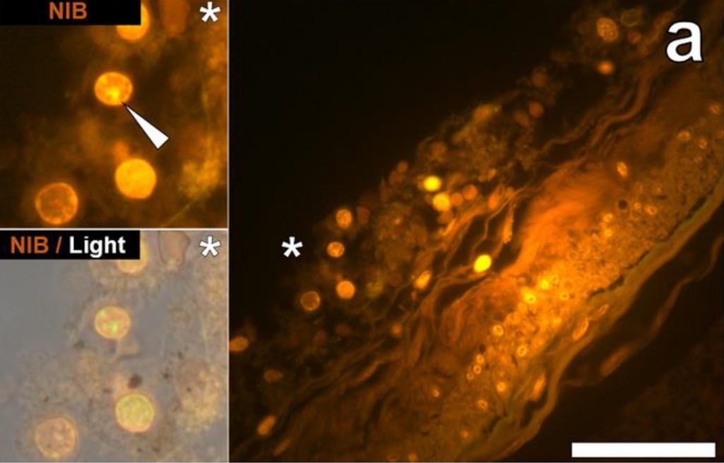
In addition to the concentration, the locations of the aluminum deposits were also examined using fluorescence microscopes. A dye that selectively stains aluminum in cells and human tissues and makes them appear orange or bright yellow was used to view aluminum on the images obtained through the microscope. Deposits were found both inside and outside brain cells. However, the most distinct observation was the presence of metal deposits in the microglia. Microglia are the main immune defense cells inside the central nervous systems and scientists concluded that the deposits seen in them were a direct indication that aluminum had somehow crossed the blood-brain barrier.

fluorescence micrograph
Figure 1 shows the cells in the hippocampus of a 50-year-old male donor used in the study by Mold et al. The white arrow indicated aluminum depositions that were observed via orange fluorescence emission. Hippocampus is the part of the brain considered to be the center of emotion and memory.

Aluminum is toxic to living cells. Although the microglia could remain functional for a certain time period, the metal will eventually show its adverse effects by disrupting this functionality. This directly correlates defective microglia with ASD. In addition to microglia, the study showed aluminum depositions in other tissues from different parts of the brain.

The study also showed great variability in the age groups of donors from 15 to 50 year olds. Initially, the high concentration seen in tissue from a 15 years old donor had greatly puzzled the researchers. However, the evidence of aluminum deposition in the microglia and other intracellular locations ties back to implicate vaccines as a potential cause of ASD and explain how such high amounts of aluminum could have deposited in the brain tissues of a 15 year old boy.

This shows the first ever instance of aluminum concentration measurement in human brain tissues from individuals with ASD. Despite the concrete results, the research was limited due to the lack of a substantial number of subjects and the minimal amount of tissue cells that could be obtained for the study. These factors render the research inadequate by itself to establish ASD as a direct outcome of aluminum deposition from vaccines in brain tissues. However, it is a major stepping stone towards realizing the potential cause of autism spectrum disorder. Now, there is a need for more research to either support or question the results of this study. 

Reference:

Mold, M., Umar, D., King, A., and Exley, C.2018. Aluminium in brain tissue in autism. Journal of Trace Elements in Medicine and Biology 46: 76-82.

Link to article

Link to feature image


Radiation in Clinical Care

Biomedical Imaging

Medical Intensive care unit (MICU) patients are exposed to radiation higher than annual US Federal occupational standard limits within a short period of time during radioscopic studies. Research conducted between January and December 2013 by scientists at the Cleveland Clinical Foundation discovered this to be true among 3% of the total 4155 patients involved in the study. 

The millisievert (mSv) is a measure of the organic effect of ionizing radiation and is known as the effective dose (ED). Statistical tests conducted on the data obtained in the study showed that 36% of the patients were exposed to radiation higher than the natural background radiation (~3 mSv). Values just 3-5 times higher have been suspected to cause carcinogenesis, although the debate is still ongoing.

Radiation is used in numerous reliable diagnostic procedures in the MICU. Although this has been overshadowing the potential risk of cancers, recently, more medical literatures are exploring its adverse effects. The study also showed an increase in cumulative ED for patients with a higher length of stay at the MICU. Among the radiation based Imaging systems used in healthcare, CT and IR caused the highest amount of radiation burden. However, diagnosis of sepsis, COPD, cirrhosis and Gastrointestinal bleeding were seen to affect cumulative ED. 

radiation burden data distribution
Figure 1 shows the distribution of data obtained by Krishnan et al for radiation burden

Ionizing radiation can lead to cancers by damaging our DNA or RNA, and cause genetic abnormalities. This is especially true for kids and young adults who have a high rate of cellular division and a longer lifespan to express the effects of radiation. Krishnan et al report that despite their adoption of the ALARA (‘as low as reasonably achievable’) principle of radiation safety, patients were exposed to substantial amounts of radiation during diagnosis. Therefore, they suggest methods for assessing the risk vs benefit of radiation therapy.

“Proactive monitoring of CED with real time display in electronic medical resorts will assist physicians in deciding the risk-benefit ratio.”

The study was limited as it was conducted among MICU patients from a single academic medical center. The estimate of ED from previous papers and the limited medical records from which data was obtained could have caused an undervaluation of the cumulative ED. Another shortcoming was the disregard for patient basic characteristics like age and sex. Nevertheless, the results denote a need to conduct more research to weigh the pros and cons of radiation in medicine and its effects on overall patient health.

This makes me wonder, are we so highly dependent on radioscopic resources that we fail to consider the harm it might be doing us? At this day and age of technological advancement, there should be active endeavors to explore the benefits and possible disadvantages of radiation in the medical industry and ways to alleviate them.

Reference: Krishnan S, Moghekar A, Duggal A, Yella J, Narechania S, Ramachandran V, Mehta A, Adhi F, Vijayan AKC, Han X, Dong F, Martin C III, Guzman J, Radiation Exposure in the Medical Intensive Care Unit- Predictors and Characteristics, CHEST (2018), doi: 10. 1016/ j.chest.2018/01/019.

Link to article

Canadian Light Source Research Office


Hormones Do It Again

T-cells

Researchers at the UPCM Sorbonne University in France have linked Autoimmune Regulator (AIRE) proteins to the high predisposition of women to autoimmune diseases (AD). Results from numerous studies correlated mutations in AIRE to the occurrence of ADs. Further research was conducted that showed a negative effect of female hormones on the functioning of AIRE.

ADs are results of abnormal immune responses caused by T cells (immune cells in the blood) that recognize body cells as foreign and attack them. AIRE are proteins present in cells of the inner lining of the thymus. They recognize such T cells and eliminate them through apoptosis (cell death) in a natural process called central immune tolerance. To back this hypothesis, research conducted on an autoimmune thyroid mouse model showed an increase in the presence of autoantibodies in the blood upon blocking AIRE expression in the thymus.

T-cells

The relationship between autoantibodies and female sex hormones is most prominent through the high incidence of ADs in females after puberty, a time of significant hormonal differences between males and females.

A study showed that the female sex hormones estrogen and progesterone caused a decrease in the expression of AIRE whereas the hormone DHT (formed from testosterone) showed an increase in expression. Estrogen has been found to contribute to this by causing hypermethylation of the AIRE gene. In this process, methyl groups are added to DNA molecules, hence changing their function without causing significant change in the structure. This is but one method by which female sex hormones lead to ADs. However, there is much more to be learned by exploring the multitude of pathways involved in the relationship between sex hormones, AIRE and ADs.

As an example, in certain body cells, AIRE has been shown to activate genes that are responsible for insulin tolerance by binding to them. Low AIRE expression has been associated with type I diabetes. Besides AIRE, there are other proteins being investigated to check for possible correlations between sex hormones and AD.

PRDM1 (PR domain zinc finger protein 1) is a protein in our body that helps prevent ADs through the deletion of defective T cells in the thymus. Research studies conducted on mice have shown gender dependence on the workings of PRDM1. However, there is no direct evidence of a connection between PRDM1 and autoimmune diseases in humans. There is much more to be explored in regard to these new results.

The reasons behind the gender based difference in AD must surely not be limited to a few proteins or sex hormones. And just how much do our hormones come into play in the determination of our susceptibility to ADs? What factors contribute to strengthen or alleviate this vulnerability? Many such questions can be raised and the answer to them all would be to dig deeper into the mysteries hidden inside of us.

Reference:

Berrih-Aknin, S., Le Panse, R., and Dragin, N.2018. AIRE: a missing link to explain female susceptibility to autoimmune diseases. Annals of the New York Academy of Sciences 1412: 21-32.

Link to article

NASA’s Marshall Space Flight Center