Molecular markers identified for autism, schizophrenia, and depression

Some psychological disorders, such as schizophrenia, tend to be highly heritable, meaning that the disorder is often passed down generationally within a family. Schizophrenia, for instance, is 60-87% heritable; if you were to have schizophrenia, there’s a 60-87% chance that one of your immediate relatives will develop symptoms, too. Similarly, major depressive disorder is 30-40% heritable. Therefore, in order to treat these disorders, its necessary to look at the genes involved. A February 2018 study published in Science found that there is significant overlap in gene expression between autism spectrum disorder, schizophrenia, and bipolar disorder, as well as an overlap between schizophrenia, bipolar disorder, and major depression. The strongest relationship was between schizophrenia and autism spectrum disorder.

Consider gene expression as a construction company. A construction company has a stockpile of materials: concrete, glass, cement, wood, nails, etc. The company has a crew of workers, and the crew is capable of building a variety of houses and apartment buildings. The construction company is analogous to the use of DNA by cells in the brain. The DNA is like the stockpile of materials. The materials are required to build anything, but the possible combinations of materials are endless. The RNA transcription mechanism in the cells is like the crew. The crew chooses which materials to use, and determines how much of each item is necessary for the project. In cells, this system is called “gene expression.” Every cell in the brain has the same DNA, or the same starting materials, but each cell has a different construction crew that decides to use the materials slightly differently; some build houses, some build apartment buildings, some build garages.

Instead of examining the DNA, or the building materials in over 700 cadaver brain samples used in the study, the researchers looked at the gene products, or what the construction crews built. It is unknown whether the gene products found in the brains caused the disorder symptoms, or gradually developed throughout life as the consequence of the disorders. But the study provides useful information regarding what proteins and structural factors manifest in disordered brains, and this information can be used to trace back to an origin point. Director of the UCLA Center for Autism Research and Treatment, and author of the study Daniel Geschwind said, “These findings provide a molecular, pathological signature of these disorders, which is a large step forward.”

The scientists found biological markers that tend to distinguish a brain with autism, for example, from the average brain. In the case of autism spectrum disorder, the study reported an increased activation of the CD11 gene, while another gene called CD2 was especially active in the brains suffering from depression. Additionally, the study mapped gene expression commonalities between brains with the same disorder, essentially establishing a molecular blueprint that can be recognized for diagnosis, and treated more effectively at the molecular level.

Sources:

Gandal, M.J., Haney, J.R., Neelroop, N.P., Leppa, V., Ramaswami, G., Hartl, C., Schork, A.J., Appadurai, V., Buil, A., Werge, T.M., Liu, C., White, K.P., CommonMind Consortium, PsychENCODE Consortium, iPSYCH-BOARD Working Group, Horvath, S., & Gerchwind, D.H. 2018. Shared molecular neuropathology across major psychiatric disorders parallels polygenic overlap. Science 359: 693–697.

Hopper, Leigh. 2018. Autism, schizophrenia, bipolar disorder share molecular traits, study finds. UCLA Newsroom. Retrieved Feb. 26 from http://newsroom.ucla.edu/releases/autism-schizophrenia-bipolar-disorder-share-molecular-traits-study-finds.

Seidel, D.C., Bulk, C., Stanley, M.A. 2017. Abnormal Psychology: A Scientist-Practitioner Approach (4th Edition). Pearson Education [print].

The Beginning of an End to the Autism-Vaccine Debate?

Autism Awareness

Autism spectrum disorder (ASD) is a developmental disorder of the nervous system. The causes of ASD are yet unknown, but it has been linked to both genetic and environmental factors. Researchers at Keele University in the UK have identified aluminum as a potential cause of autism based on a study conducted in 2017 on brain tissue from people diagnosed with ASD.

Aluminum is used in vaccines to enhance the body’s immune response to antigens (harmful or toxic substances). The vaccine-autism debate is highly controversial, but animal models have linked the use of aluminum in vaccines to ASD. The results of this study on human cells further assert these findings.

Aluminum content was measured in 0.3g tissue samples from different regions of the brain of 5 individuals using atomic absorption spectrometry. This method utilizes the difference in the light absorption capabilities of different atoms to find the chemical composition of samples. The values ranged from 1.20 to 4.77μg/g. Past studies have suggested values ≥2.00μg/g as pathologically concerning and those ≥3.00μg/g as pathologically significant. The results showed at least one tissue in each individual that exceeded the established pathologically significant value.

Some of the values recorded were the highest ever measured (17.10, 18.57 and 22.11μg/g).

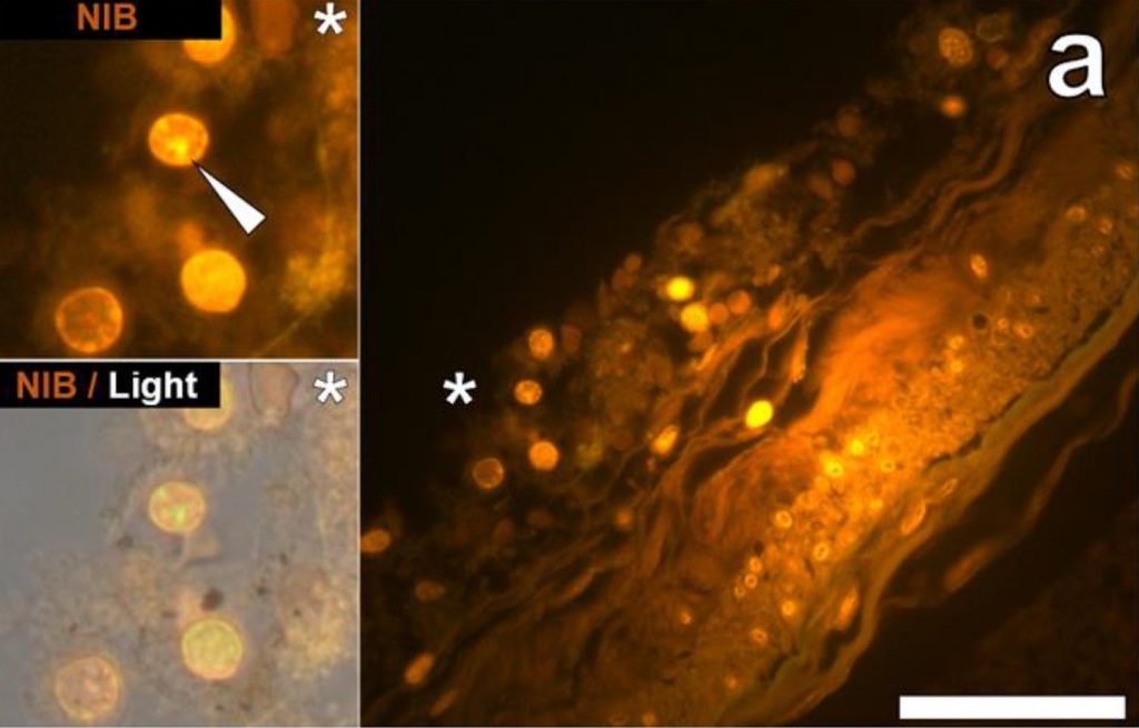
In addition to the concentration, the locations of the aluminum deposits were also examined using fluorescence microscopes. A dye that selectively stains aluminum in cells and human tissues and makes them appear orange or bright yellow was used to view aluminum on the images obtained through the microscope. Deposits were found both inside and outside brain cells. However, the most distinct observation was the presence of metal deposits in the microglia. Microglia are the main immune defense cells inside the central nervous systems and scientists concluded that the deposits seen in them were a direct indication that aluminum had somehow crossed the blood-brain barrier.

fluorescence micrograph
Figure 1 shows the cells in the hippocampus of a 50-year-old male donor used in the study by Mold et al. The white arrow indicated aluminum depositions that were observed via orange fluorescence emission. Hippocampus is the part of the brain considered to be the center of emotion and memory.

Aluminum is toxic to living cells. Although the microglia could remain functional for a certain time period, the metal will eventually show its adverse effects by disrupting this functionality. This directly correlates defective microglia with ASD. In addition to microglia, the study showed aluminum depositions in other tissues from different parts of the brain.

The study also showed great variability in the age groups of donors from 15 to 50 year olds. Initially, the high concentration seen in tissue from a 15 years old donor had greatly puzzled the researchers. However, the evidence of aluminum deposition in the microglia and other intracellular locations ties back to implicate vaccines as a potential cause of ASD and explain how such high amounts of aluminum could have deposited in the brain tissues of a 15 year old boy.

This shows the first ever instance of aluminum concentration measurement in human brain tissues from individuals with ASD. Despite the concrete results, the research was limited due to the lack of a substantial number of subjects and the minimal amount of tissue cells that could be obtained for the study. These factors render the research inadequate by itself to establish ASD as a direct outcome of aluminum deposition from vaccines in brain tissues. However, it is a major stepping stone towards realizing the potential cause of autism spectrum disorder. Now, there is a need for more research to either support or question the results of this study. 

Reference:

Mold, M., Umar, D., King, A., and Exley, C.2018. Aluminium in brain tissue in autism. Journal of Trace Elements in Medicine and Biology 46: 76-82.

Link to article

Link to feature image