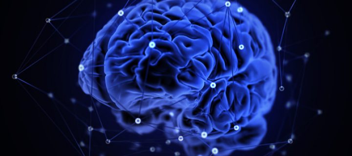
There are so many different types of cancer that exist in all parts of our body, and each subset of cancer has its own challenges during treatment. Brain cancer in particular can be challenging to treat. For patients with a brain tumor, the first line of treatment is often surgery to remove as much of the tumor mass as possible. While the aim is to remove enough of the tumor so that patients have a reasonable chance of survival, surgeons need to diagnose exactly where the tumor is located so that they don’t remove too much healthy tissue, which would have harmful consequences such as memory loss, vision loss or loss of the ability to move our muscles. Currently, trained pathologists stain the tissue with special dye and then the pathologists analyze the results and report to the surgeon their diagnosis. As you can imagine, this process of staining the tissue and then confidently reporting where the tumor is located can be time-consuming and requires trained pathologists to always be available. Therefore, researchers and surgeons at the University of Michigan collaborated to identify a ‘new workforce’ to aid in this process.
The research team wanted to test if they could combine an imaging technology (called SRH) with artificial intelligence. It was THE dream team! SRH is a
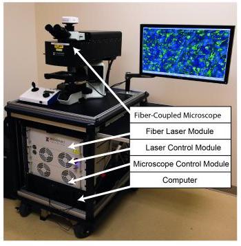
The microscope is used to capture images of the tissue samples from the brain. The computer analyzes the image and produces a diagnosis.
specialized form of microscopy that can be used to identify tissue samples in the operating room in a similar way that the pathologists stain tissues to identify them. This imaging technology was then connected to a computer equipped with the ability to perform tasks at the level of human intelligence. Otherwise known as Artificial Intelligence. The AI computer is ‘trained’ to recognize patterns and perform tasks, such as analyzing the images created by the SRH microscopy.
With the concept developed, the researchers began training the computer to recognize tissue samples as tumor tissues or normal tissues. They used over 2.5 million tissue samples (a very rigorous training indeed!). With the computer well trained, the researchers enrolled 280 patients in a clinical trial where half of the patients were diagnosed with the conventional method (pathologists who stain and analyze the tissues), and the other half with the new technology (images diagnosed by the AI machine).
Amazingly, the Artificial Intelligence technology correctly diagnosed 94.6% of the time, while conventional pathologist-based analysis had an overall accuracy rate of 93.9%.
Interestingly enough, in the 5% of cases where the AI technology incorrectly classified the tumors, the pathologists made the correct diagnosis. Additionally, each time the pathologists were incorrect, the AI system had made the correct diagnosis. This ability for the two diagnosis methods to cross check one another suggests the need for AI technology to support the work of pathologists.
In addition to accurate diagnosis, the AI technology also reduces diagnostic time significantly from 30 minutes to 3 minutes.
This new technology could help transform the field of cancer surgery as it will allow doctors to make the most accurate decision about a treatment course. The extent of tumor removal, that is how much of the tumor is actually removed, dictates the outcome for patients. The use of this technology could help to remove as much tissue as possible, which prolongs patients’ lives, while not damaging all the healthy tissue that is essential for us to live healthily.
This research is just one example of the remarkable interdisciplinary nature of science and how collaborating across fields such as computer science, medicine and cancer research in this case, can lead to positive impacts on many people’s lives.
For more information:
Orringer, D., Pandian, B., Niknafs, Y. et al. Rapid intraoperative histology of unprocessed surgical specimens via fibre-laser-based stimulated Raman scattering microscopy. Nat Biomed Eng 1, 0027 (2017). https://doi.org/10.1038/s41551-016-0027.
Alex
"This title was very eye catching! That is so interesting that such a ..."
Alex
"This is really interesting! The fact that crops and plants are damaged is ..."
Alex
"Well done, this article is great and the information is very captivating! Ethics ..."
Alex
"I was intrigued throughout the whole article! This is such an interesting topic, ..."
Alex
"This is such an interesting article, and very relevant!! Great job at explaining ..."
Grandpa
"Honey You Did a good job I will forward to my eye doctor "
murphymv
"This article is fascinating because it delves into the details of the research ..."
murphymv
"I agree, adding the photo helped solidify the main finding. "
murphymv
"This is a fascinating finding. I hope this innovative approach to improving transplants ..."
Sherzilla
"This is a great article! I would really love to hear how exactly ..."
Sherzilla
"It's disappointment that these treatments were not very effective but hopefully other researchers ..."
Sherzilla
"I agree with your idea that we need to shift our focus to ..."
Sherzilla
"It's amazing to see how such an everyday household product such as ..."
Lauren Kageler
"I will be interested to see what the data looks like from the ..."
Lauren Kageler
"A very interesting article that emphasizes one of the many benefits that the ..."
maricha
"Great post! I had known about the plight of Little Browns, but I ..."
Sherzilla
"I assumed cancer patients were more at risk to the virus but I ..."
Sherzilla
"Great article! It sheds light on a topic that everyone is curious about. ..."
maricha
"This article is full of really important and relevant information! I really liked ..."
maricha
"Definitely a very newsworthy article! Nice job explaining the structure of the virus ..."
maricha
"It's interesting to think that humans aren't only species dealing with the global ..."
murphymv
"This is very interesting and well explained. I am not too familiar with ..."
Lauren Kageler
"Great article! This post is sure to be a useful resource for any ..."
Lauren Kageler
"Definitely seems like an odd pairing at first, but any step forward in ..."
murphymv
"What an interesting article! As you say, height and dementia seem unrelated at ..."
murphymv
"Great article! I learned several new methods of wildlife tracking. This seems like ..."
murphymv
"Very interesting topic! You explained cascade testing and its importance very well. I ..."
Alex
"This article is really interesting! What got me hooked right away was the ..."
Sabrina
"I found this article super interesting! It’s crazy how everyday products can cause ..."
Erin Heeschen
"I love the layout of this article; it's very eyecatching! The advancements of prosthetics ..."
murphymv
"Awesome article! I like the personality in the writing. Flash Graphene not only ..."
murphymv
"Very interesting work! I don't know a whole lot about genetics, but this ..."
Cami Meckley
"I think the idea of using virtual reality technology to better help prepare ..."
Erin Heeschen
"I wonder if there's a connection between tourist season and wildfires in the ..."
Ralph berezan
"Not bad Good work "
Michelle
"This sounds like it would be a great tool for medical students! ..."