Change Your Mind: How Stress Reshapes Your Brain

The hippocampus in a human brain.

By Kelly Lohr

It has been known for a while that too much stress can be bad for your health.  A new study now shows that it can affect your brain too.  Research through a collaboration between Rockefeller University and Cornell University suggests that stress can been linked to harmful changes in some brain structures.  Sometimes these brain changes can be advantageous, such as making new synaptic connections to remember and learn from a stressful, life-threatening event.  However, some changes can  be detrimental.

A mouse hippocampus labeled with NeuroTrace® green fluorescent Nissl stain

The project has identified a protein possibly involved in remodeling the brain under stress.  It was found that the brains of mice lacking the protein called brain-derived neurotrophic factor (BDNF) look like the brains of stressed mice.  The study examined changes in the neurons of the hippocampus, a brain area important in memory, mood, and cognition.  When normal mice were stressed through confinement to a small space, the tiny projections on their neurons called dendrites retracted in the hippocampus.  The hippocampus itself was also reduced in overall volume.  The study compared these mice to other mice that were missing a copy of the gene that produces BDNF.  It was found that these genetically-altered mice had brains resembling those of stressed mice.

Not only does this finding show that stress can produce brain changes.  Bruce McEwan of Rockefeller University suggested that BDNF also may be “one of the proteins that play a role in mediating the brain’s plasticity.” This holds promise for a better understanding of the role of neuronal remodeling in the hippocampus and its importance in memory and emotion.

Written April 13, 2010

For more information, visit http://www3.interscience.wiley.com/journal/123249229/abstract?CRETRY=1&SRETRY=0.

History of Smoking Reduces Risk of Parkinson’s Disease

By Kelly Lohr

Believe it or not, a new study has shown that a history of cigarette smoking may actually benefit your health.  Over the last decade, Honglei Chen led a study out of the National Institute of Environmental Health Sciences in Research Triangle Park, North Carolina examining long-term health effects of the habit.  Over 300,000 AARP members between the ages of 50 to 71 were surveyed about lifestyle choices over a ten-year period.  Of these subjects, 0.05% of the individuals developed Parkinson’s disease.

Parkinson’s disease is a neurodegenerative disorder characterized by the breakdown of cells which release the neurotransmitter dopamine in a brain area known as the substantia nigra.   Typical symptoms of Parkinson’s include uncontrollable muscle movements, poor posture, and rigidity.  Of the participants from Chen’s study, it was found that current smokers reduced their risk of Parkinson’s disease by 44% as compared to non-smokers.  Previous smokers who had quit reduced their risk of Parkinson’s by 22%.

Interestingly, the risk of developing Parkinson’s disease did not change based on how many cigarettes a person smoked per day.  Instead, the length of the history of smoking was correlated to reduction in disease risk.  Those who smoked for at least 40 years were 46% less likely to develop Parkinson’s, whereas those who smoked between 30 and 39 years reduced their risk by 35%.  However, individuals who smoked for nine years or less only reduced their risk by 8%.

Despite Chen’s findings, smoking does not slow the progression of Parkinson’s once it develops.  For this reason, experts do not suggest that nicotine or other chemicals in cigarettes should be considered as effective Parkinson’s disease treatments.  Despite this, an improved understanding of the mechanisms behind the reduced risk may lead to breakthroughs in the causes of the disease.

For more information, visit http://www.neurology.org/cgi/content/abstract/74/11/878.

The Sea Squirt: An Answer to Alzheimer’s?

Ciona intestinalis

By Kelly Lohr

The newest breakthrough in Alzheimer’s research is coming from an unlikely source–a sea squirt.  Just this week (March 2, 2010) Mike Virata and Bob Zeller of San Diego State University believe that Ciona intestinalis, known commonly as the sea squirt, may be the perfect model organism for this disease.

The brains of Alzheimer’s patients are typically filled with tangles and plaques made of the protein fragment beta-amyloid.  Alzheimer’s disease affects nearly 4 million Americans and an estimated 27 million people worldwide. It is the most common form of age-related dementia and has no cure. Current drug regimens only relieve symptoms and cannot halt the progression of the disease. Research in the scientific community is currently  aimed at slowing the disease through drugs such as Aricept and Namenda which are focused on decreasing plaque accumulation.

Recently, research has shown the need for an improved model organism to aid  in understanding the pathology of the disease.  Currently, genetically modified strains of mice have been the organism of choice in the research of this disease. However, there are limitations in the use of mice including an extremely long waiting period for plaque development like those seen in Alzheimer’s brains. Also, these mice do not contain the same genetic mutations linked to hereditary risk of Alzheimer’s disease.  Mice are also more costly to purchase and maintain for research.

Sea squirts are tunicates, marine organisms with a hard outer tunic and a soft body. They live on underwater structures and are filter feeders that eat small plant material. It has been suggested that sea squirts are actually our closest invertebrate relatives.  As far as research benefits, sea squirts share nearly 80% of our genes and resemble vertebrates in their immature form.  These animals are inexpensive to house and contain all of the genes needed for the development of Alzheimer’s plaques in humans.

An immature sea squirt.

Virata and Zeller found that by giving the immature sea squirt amyloid precursor protein, a mutant protein linked to hereditary Alzheimer’s, sea squirts developed brain plaques in a single day.  Further, these plaques and the behavioral deficits seen in these animals were able to be reversed using a drug meant to remove plaques.  Such techniques have been ineffective in all other invertebrate models, including the commonly used nematode, C. elegans.  Now, investigators can be freed from genetic, time, and financial constraints.  These findings provide a resource for an entirely new take on Alzheimer’s research…all because of a sea squirt.

For more information, click here.

Got milk? Vitamin D, a key player in multiple sclerosis

By Kelly Lohr

Drinking milk may do a lot more than just strengthen our bones. A study out of the Harvard School of Public Health in Boston recently suggested drinking milk during pregnancy may markedly reduce the chance of the child developing multiple sclerosis (MS) later in life.

Vitamin D can be found in dairy, fatty fish, and supplements. Exposure to sunlight also produces this helpful vitamin.

Lead by Fariba Mirzaei, MD, the study examined over 35,000 female nurses whose mothers had completed questionnaires recording their diets during their pregnancies with their now-grown daughters.  The work occurred over a 16-year period, during which 199 women developed MS.

MS is a degenerative disease that attacks the central nervous system (CNS), including the brain, spinal cord, and optic nerves. The symptoms vary, ranging from numbness in the arms and legs to paralysis and loss of vision. Unfortunately for its sufferers, the progress and severity of MS are often unpredictable. The neurons in our body are partially covered in a fatty substance called myelin in order to insulates the cells and  to allow them to transmit signals quickly. If the myelin is damaged, these signals can be delayed. MS results in the destruction of this insulating myelin in the CNS. This breakdown is thought to be caused by the body’s immune system attacking the myelin sheath.

Normal and damaged myelin in neurons.

The researchers lead by Dr. Mirzaei found that the risk of MS was lower in women whose mothers had high milk or vitamin D diets during pregnancy. Women whose mothers drank four glasses of milk per day had a 56% less chance of developing MS than those whose mothers drank less than three glasses per month. In general, women in the top 20% of vitamin D intake had a 45% less chance of having a child develop MS than those in the bottom 20% of vitamin D intake.

Vitamin D can come in many forms including fatty fish, milk and dairy products, and exposure to sunlight. Supplements could also be used to counter vitamin deficits in the diet. This study serves as evidence of a growing role for vitamin D in the pathology of MS. Prevention may play an important part in the disease, perhaps starting as early as pregnancy.

Source: http://www.medscape.com/viewarticle/716913

Making new connections: Stem cells as treatment for ALS

By Kelly Lohr

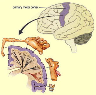
The motor cortex in the human brain is mapped to match specific body parts. Body parts with more devoted cortex area are generally more sensitive or have finer motor control.

              Imagine slowly losing control of your muscles, first with a few twitches in your arms and legs or a slurred word here or there. Muscle failure will continue until it eventually stops your ability to move, speak, and breathe.  This is the life of a patient suffering from amyotrophic lateral sclerosis (ALS) also known as Lou Gehrig’s disease, a progressive neurodegenerative disorder.  Currently, there is little treatment for the rapid course of this disease, but James Weimann, PhD, of Stanford Medical School provides a new hope.

            Weimann is part of a team of neuroscientists using transplanted neurons grown from embryonic stem cells to replace damaged cells in young animals.  This finding is the first of its kind in that the stem cells can be directed to take on the jobs of specific brain cells while also making the correct connections with other cells. Weimann’s cells transmit information from the cortex, the neural tissue that is outermost part the mammalian brain, specifically areas needed for motor function.

             Up until this point, the issue of stem cell transplantation in the brain was making the proper neuronal connections.  As an adult organism, creating the accurate connections in the nervous is extremely complex.  During development, superfluous neural connections deteriorate with lack of use. Only the pathways with the most activity remain in adulthood.  The chemical or physical signals that once lead the way in development are no longer present.  Without such cues, it is difficult for neurons to reach their target areas. For example, the stem cells created in Weimann’s lab must make connections with motor cortex in order to be an effective treatment for disorders like ALS or a traumatic brain injury.  Incorrect connections could result in further erratic brain function.

A step in the processing of human embryonic stem cells.

           While Weimann’s work holds a lot of potential for further progress and treatments, the studies have involved transplantation in young animal models.  Since the majority of neurodegeneration takes place in older adults, the next step will be to explore stem cell transplantation in adult animals.  Weimann and his team are hopeful that these newest findings will soon be used in treatment of neurons that are lost or damaged due to spinal cord injuries or diseases like ALS.