Researchers solve viral superinfection mystery

By Liz H. ‘10

Interaction between a T-cell (purple) and another cell of the immune system.

The ability of a common virus known as CMV to cause a “superinfection” and infect humans multiple times has puzzled scientists until recently.  Researchers at the Oregon Health and Science University Vaccine and Gene Therapy Institute have reported the mechanism that CMV uses to evade the immune system and re-infect humans, in a study published in the April 2nd issue of Science.  Their findings shed light on how this virus may be used in the development of CMV-based vaccines for other diseases.

Cytomegalovirus (CMV) does something that not many viruses can do:  it can re-infect people who previously have been infected by CMV and have already developed an immune response to the virus.  This is unusual for a virus, because the immune system usually “remembers” previous infections with viruses and other pathogens and can mount a strong immune response upon re-infection with a specific pathogen.

The researchers studied CMV-infected monkeys in order to understand how the virus overcomes detection by the immune system.  They discovered that CMV avoids a special type of white blood cell called CD8+ T cells, which are responsible for killing cells that are infected with a pathogen.  CD8+ T cells recognize infected cells by small molecules on the exterior surface of infected cells known as MHC I.  These molecules display small pieces of an invading pathogen and present them to the T cells.  This presentation of infectious material ultimately signals T cells to start destroying infected cells.

In order to evade these T cells, CMV makes proteins that interferes with the presentation of viral pieces by MHC I molecules and stops them from recruiting T cells to infected cells.  “In essence, CMV is able to cut off an infected cell’s call for elimination. This allows CMV to overcome this critical immune barrier during re-infection,” explains author Klaus Frueh.

The study has interesting implications in the design of CMV-based viral vaccine vectors.  Viral vaccine vectors contain a modified, harmless virus that carries a vaccine for another pathogen to the body.  Although the body develops immunity to the pathogen, it also develops immunity to the viral vector, which means that the viral vector can only be used for one type of vaccine.  Since CMV does not elicit an immune response upon re-infection, it makes an attractive vaccine vector candidate that could potentially carry vaccines against other pathogens, such as HIV, hepatitis C, tuberculosis, and malaria.

CMV is a member of the herpesvirus family and infects 50-80% of the US population by age 40.  Most people do not have any symptoms of CMV infection and do not become ill.  But for those with weakened immune systems, including infants and the immunocompromised, CMV can cause serious complications.  With this new understanding of how CMV evades the immune system, scientists may be able to start utilizing the virus for the benefit of human health.

Want more information?

CMV infection

Press Release

An uncommon use for a common drug offers hope to millions of HIV-positive patients

By Liz H. ‘10

Microscopic view of HIV (green) emerging from an infected T-cell. CDC

A promising new HIV treatment has been discovered in an unlikely source:  a widely available acne medication developed in the 1970s.  A team of scientists from Johns Hopkins University reports that minocyclin stops HIV-infected human cells from reactivating and replicating, in a study published in the April 15th issue of the Journal of Infectious Diseases.  Their findings may lead to an improved and more effective treatment regimen for HIV infection.

The researchers focused their study on latent, non-replicating HIV-infected human T-cells.  T-cells are a type white blood cell that normally fights infection.  HIV infects T-cells and can “rest” inside of them for an extended period of time.  The virus does not harm the T-cell during this latent phase, but can eventually “wake-up” and re-activate the T-cell, which spreads HIV infection and weakens the immune system.

In this study, the scientists treated latent HIV-infected human T-cells with minocycline and measured the level of re-activated T-cells over time.  They also performed the same measurements on cells that were not treated with minocycline.  The researchers found that the minocycline-treated cells did not display detectable levels of reactivation while the untreated cells displayed elevated levels.

Upon closer analysis of the activity of minocycline inside of cells, the scientists discovered that the drug interferes with important cellular communication pathways that cause the T-cell to activate and spread HIV to other cells.  “It prevents the virus from escaping in the one in a million cells in which it lays dormant in a person…That’s the goal:  Sustaining a latent non-infectious state,” explains Gregory Szeto, a Hopkins graduate student who worked on the project.

These findings suggest that minocycline could be used in conjunction with HAART, the current HIV treatment standard, to keep the virus dormant inside of T-cells.  “While HAART is really effective in keeping down active replication, minocycline is another arm of defense against the virus,” says author Janice Clements.  Minocycline is an attractive addition to the current arsenal of HIV medications because it is relatively inexpensive, does not inhibit the ability of T-cells to fight other infections, and is not likely to cause viral drug resistance.

Current treatment for HIV/AIDS involves a combination therapy approach known as HAART.  Patients on HAART take at least 3 antiretroviral drugs daily that act on the virus in different ways to reduce its levels in the bloodstream.  Although HAART can extend the life of an infected individual, it is not a cure and causes unpleasant side effects and the development of drug resistance.  For the 40 million HIV-positive individuals worldwide, this new use for minocycline promises improved outcomes.

Want more information?

What is HIV?

Press Release

Bird flu: A Thing of the Past?

By Nick Gubitosi

This past Friday (February 26, 2010) a group of scientists led by a virologist from the University of Wisconsin published a study about a new antiviral, which was found to be highly effective against the pathogenic H5N1 avian influenza virus.  What stands out about this new antiviral, known as CS-8958, is that it has been proven to be effective against Tamiflu resistant strains of H5N1. This makes it a promising candidate for the future treatment and prevention of the bird flu.

Antiviral drugs are used in the treatment of viral infections by inhibiting the development of disease causing pathogens, and are a vital component in the countermeasure against human influenza viruses.  Recently many new strains have been emerging, which show resistance to Tamiflu, an antiviral that slows the spread of the influenza virus within the body.  These resistant strains pose a threat and make the development of new antivirals a pressing issue.

Professor Yoshihiro Kawaoka from the University of Wisconsin and his team of scientists tested a drug created from a novel neuraminidase inhibitor on mice in order to see its effectiveness against H5N1 strains of influenza.  Neuraminidase inhibitors are a class of antiviral drugs that specifically target the influenza virus by blocking one of its proteins, therefore preventing its replication within the body.

They began their tests by giving a single dose of the CS-8958 antiviral drug nasally to mice, two hours after infection with the H5N1 influenza virus.  The results showed that the survival rates were higher in the mice given this new drug when compared to mice given a standard five day treatment with Tamiflu.  In another experiment, CS-8958 was found to be effective against highly pathogenic and Tamiflu resistant strains of H5N1, while it was also shown to protect mice against lethal H5N1 infection when it was administered seven days before infection with the virus.

With the information gained from this study, future treatment and prevention of H5N1 with this CS-8958 antiviral could be the most effective treatment to date due to its ability to eliminate newly emerging drug resistant strains in only one dose.  While future studies still need to be conducted to make sure that these results are the same when tested on humans, the potential of this new antiviral is promising and could possibly put an end to the fear of the bird flu pandemic.

Click here for source

Taking the flight (and bite) out of the pesky mosquito

By Liz H.

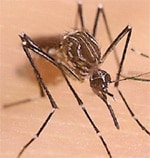
The bite of the female Aedes aegypti mosquito can transmit the virus that causes Dengue fever to humans
The bite of the female Aedes aegypti mosquito can transmit the virus that causes Dengue fever to humans.

The days of the flying mosquito may be drawing to a close.  In a study published in the February 22nd issue of the Proceedings of the National Academy of Science, a team of American and British researchers report that they have engineered a mosquito in the lab that produces offspring that cannot fly and consequently cannot infect humans with the virus that causes Dengue fever (full article).  Their findings may lead to a sustainable mosquito population suppression strategy that dramatically reduces human morbidity and mortality from a variety of diseases transmitted by mosquitoes.

In this study, the scientists specifically focused on the Aedes aegypti mosquito that causes Dengue fever.  The researchers manipulated the genetic material of the males of this species in the lab to carry a novel trait:  the inability to fly.  When these modified males were mated with normal, wild-type females, they passed the trait on to their female offspring.  By rendering the female offspring flightless, the scientists effectively imposed a death sentence on this group.  If the females cannot fly, they cannot elude predators, mate with males, escape from water, or seek out human blood.  Most importantly a flightless female may lead to the eradication of Dengue fever, since the disease is transmitted by the bite of female Aedes aegypti mosquitos.

The researchers predict that 6-9 months after introducing the modified males into the wild, the wild-type females in the area will be completely replaced by the flightless offspring of the modified males.  This is big news with important applications in the control of mosquito-borne disease.  This method of control offers several advantages over traditional techniques because it specifically targets the species of mosquito that causes Dengue fever and bypasses the use of toxic insecticides.  And as senior author Luke Alphey notes, “Another attractive feature of this method is that it’s egalitarian: all people in the treated areas are equally protected, regardless of their wealth, power or education.”

The next step for the researchers is to study the mating competitiveness of the modified males in the wild and whether their flightless female offspring will actually suppress the wild-type population as predicted.  Additionally, the methods used by these scientists could be adopted to control other species of mosquitoes that spread serious diseases such as West Nile virus and malaria.

Dengue fever is a flu-like illness with no vaccine or treatment that infects 50-100 million people each year in over 100 countries in tropical and subtropical climates, including Puerto Rico and tourist destinations in Latin America and Southeast Asia.  It is the most common mosquito-borne disease and the CDC estimates that one third of the world’s population lives in areas where the disease is endemic.  Other diseases transmitted by mosquitoes include West Nile virus, malaria, Rift Valley Fever, and Yellow fever.  Taken together, these illnesses represent growing public health issues that require effective and sustainable mosquito population control measures.  The flightless mosquito may just be the answer to this urgent problem.

Want more information?

Press release

CDC site on Dengue fever

Flu to the rescue!

By Shelly Hwang

February 17, 2010

It’s flu season, and with the H1N1 virus being the spotlight of current news and the CDC pushing for nation-wide flu vaccination, people have become terrified of the influenza virus. However, a recent study done by researchers at the Yale University School of Medicine (published in the February 18 issue of Cell Host and Microbe) revealed that the stress response caused by the flu actually protects against death by secondary infection by using mice with bacterial infections.

Influenza can damage the lungs but usually does not kill. However, secondary infections such as pneumonia can occur after infection with the influenza virus and are much more deadly. Each year, more than 200,000 U.S. residents are hospitalized for flu-related complications, and about 36,000 Americans die on average per year from complications of the flu (CDC Statistics).

While previous studies on the flu have shown repressing of the immune system, such studies have only studied a single pathogen and focused on local effects of influenza at the site of infection. In reality, organisms are exposed to multiple infectious agents at a time and the effect of influenza on the whole immune system has not been studied.

This study, led by Dr. Rusian Medzhitov from the Department of Immunology, used a mouse model to examine the effects of the lung infection caused by influenza on the immune response to bacterial infection. Surprisingly, the researchers found that the influenza lung infection led to increased production of glucocorticoids (GC), which are produced in response to stress and known to play a key role in regulating inflammation. They found that virus-induced GC production is essential to controlling inflammation, as shown by the death of mice lacking GC’s that were infected by multiple pathogens.

So the next time you find yourself miserable and overwhelmed with the unpleasant flu FACTS symptoms (Fever, Aches, Chills, Tiredness, Sudden symptoms), remember to thank the virus for protecting you from fatal secondary infections.

Press Release

Department of Immunobiology at Yale

Bullet shaped virus may be the future of new cancer and HIV treatments

By Liz Humes

An obscure virus that does not harm human cells has been generating a wave of excitement in the scientific community.  So what is the big deal?  A team of researchers from UCLA has reported the 3D structure of the vesicular stomatitis virus (VSV) in a break-through study published in the February 5th edition of Science (full article).  Their findings may shed light on how VSV can be manipulated and used in the treatment of cancer and in the development of vaccines for HIV and other harmful viruses.

The researchers used advanced, cutting-edge imaging techniques to visualize the 3D structure of VSV, which appears to have a bullet shaped head and cylindrical trunk.  They also characterized how the virus comes to form this bullet shape.  With this additional level of understanding of the physical structure of the virus, scientists believe that they can find ways to modify the structure of the virus and use it to treat and prevent illnesses such as cancer and AIDS.

As author Z. Hong Zhou remarked, “This work moves our understanding of the biology of this large and medically important class of viruses ahead in a dramatic way.”

VSV is a model virus that scientists use in the laboratory to study dangerous viruses that cause illnesses such as the flu, measles, and rabies.  Previous studies have shown that VSV can detect and kill human cancer cells.  Other studies have addressed the question of how to manipulate the virus to deliver a vaccine against HIV to the human body.

3D animation of VSV trunk

(Video is a 3D animation of the lower trunk structure of VSV-source)

A closer look at vaccine technology

A current trend in vaccine development is to use harmless viruses as “vectors” that can carry a specific vaccine to human cells.  These viruses have been engineered in the laboratory to carry pieces of genetic material from other pathogens and when they attach to human cells, they inject this genetic material into the cells.  These actions mirror an infection by the pathogen itself, although the virus vector does not actually cause an infection, and stimulates an immune response.  The human body then remembers how to respond to this pathogen the next time it encounters the pathogen and the body is protected from infection.

Modified versions of the viruses that cause the common cold and small pox are being studied in addition to VSV for use as vaccine vectors.  Given the potential that this type of vaccination has to prevent deadly infections from viruses and bacteria, this is an area of research one should surely keep an eye on.

Want more information?

Press release

Vaccination information

HIV vaccination development

Newly discovered antiviral fights HIV, Ebola and other deadly viruses

By Nick Gubitosi            February 4, 2010

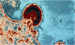
A picture of the HIV virus attacking a lymphocyte

A group of researchers lead by scientists from UCLA have identified a “broad spectrum” antiviral small molecule which targets the many envelope encased deadly viruses that exist today.  This antiviral would fight enveloped viruses such as HIV, Ebola, and influenza, as well as viruses that haven’t even been discovered yet.

Dr. Benhur Lee, an associate professor at UCLA, was working with colleagues on 23 various pathogens when they discovered that this antiviral molecule, known as LJ001, only interfered with enveloped viruses through a mechanism which is still not fully understood.

This LJ001 molecule binds to both healthy and viral cells within the body, but only causes harm to the viral cells.  Unlike the healthy cells in your body, viral cells lack the ability to repair themselves because they are not metabolically active.  Therefore the damage done to the viral cells is permanent, while it is completely harmless to the healthy body cells.

Broad spectrum antivirals are hard to find, and usually accompanied with many shortcomings.  One such antiviral, Ribavirin, targets RNA replication and is only effective against a few viruses, is too expensive for widespread use, and produces unwanted side effects.  LJ001 targets viral structure, does not appear to be toxic, and can attack a large group of viruses, making LJ001 the first antiviral of its kind.

Viruses can differ from one another and even mutate as seen with HIV, making them extremely hard to fight off.  Using an antiviral such as LJ001, which safely targets a feature common to an entire class of viruses, may be the potential answer to this problem.

See here for Press Release

Not Your Average Fairytale

By Kristen Kocher                        February 4, 2010

Numerous genetic diseases, especially hereditary brain diseases, are untreatable therefore subjecting many individuals to a life of endless pain and suffering. However, in recent years with the development of the technique of gene therapy, new hope has been brought to life in those diagnosed as “terminally ill” with the promise of the “happily ever after” ending that everyone deserves.

Gene therapy is still not used as a mainstream medical technique because much of the process is still in the developmental stages. Recently, geneticists have been desperately working to perfect the successful transport of therapy genes into brain cells. In many cases, the diseases are caused by a single gene or protein mutation but can cause devastating affects, which normally result in the loss of brain cells and fatality.

A recent scientific breakthrough has finally made it possible for therapy genes to be inserted into brain cells and cure certain genetic diseases.  Before this discovery, therapy genes were only administered through the use of viruses, predominantly the herpes virus, HSV-1. While HSV-1 has the ability to effectively transport large genes into the nucleus of the targeted cells, once the genetic information enters the nucleus it is unable to be integrated into the mammalian, host genome. This proves to be unhelpful as the therapeutic information is quickly silenced and within a few days the effects of gene therapy are no longer visible.  

Another molecule used for gene therapy transport is known as “Sleeping Beauty”. The aforementioned molecule is named as such because it is innately a silent gene that was activated, or “awakened”, by scientists. The discovery of this molecule is beneficial because it has the ability to take the target gene intended for therapy into the nucleus and integrate it directly into the mammalian genome.  The genes transported by Sleeping Beauty, however, must be relatively small, roughly 15 to 30 times less than the amount of DNA carried by HSV-1. This is unfortunate because the genes that are used for treatment of diseased brain cells are predominately large and cannot be carried by Sleeping Beauty.

So, where does the happy ending come in? These two molecules individually have characteristics that make them useful in therapy gene transport but separately cannot aid in the treatment of brain disease. However, thanks to the research of William Bowers, Ph.D. and graduate student Suresh de Silva, this blockade has been removed. With the creation of a hybrid molecule made up of both HSV-1 and Sleeping Beauty, geneticists have been able to successfully integrate large therapy genes into the mammalian genome, which, though current experiments, have resulted in long-term therapeutic gene expression. The creation of this hybrid therapy gene transport molecule promises a bright future and “happy ending” for those suffering from terminal, genetic disease.

Original Press Release

Find out more about the projects going on in Bowers Laboratory