Discoveries in Diabetes, Depression, and Dementia

By Shelly Hwang

Diabetes, Depression, and Dementia are three of the most common medical conditions among Americans today. A recent study released on March 5 by a group of researchers at the University of Washington (UW) revealed that depression in diabetic patients doubles the risk of developing dementia, a finding that may affect the way that depression is screened and treated in order to prevent the development of other diseases.

Dementia is the gradual loss of cognitive and reasoning abilities, including memory loss, wandering, inability to do basic math, and forgetting familiar things or people.  Depression is a mental disorder marked by low mood and poor concentration. Diabetes is a medical condition in which a person has a high blood sugar level. While both diabetes and major depression have been shown to be separate risk factors for dementia, the effect of both diabetes and depression on dementia has not been studied. It turns out that adults with both conditions are twice as likely to develop dementia, compared with adults with only diabetes.

This project, led by Dr. Wayne Katon, a professor of psychiatry and behavior sciences at UW, is a part of the Pathways Epidemiological Follow-Up study, which examines adults from the Group Health Cooperative’s diabetes registry. Patients from nine clinics in western Washington State were studied for five years. 163 of 3,382 (4.8%) patients with diabetes alone developed dementia, while 36 of 455 (7.9%) of the diabetes patients with major depression were diagnosed with dementia. This presents a 2.7 fold increase of dementia in diabetic patients with depression.

Depression is common among individuals with diabetes. Previous studies found depression increases mortality rate among diabetes patients, in addition to health complications. However, the way the two diseases interact is unknown. Perhaps they interact by genetics, increasing stress levels, or resulting in unhealthy behaviors such as smoking, lack of exercise, and over-eating, which raise the risk of dementia. Diabetes is a known risk factor for dementia because it causes blood vessel problems, tissue damage, and increased blood sugar levels, which all increase odds of developing dementia. Although the link between depression, diabetes, and dementia is still not understood, it is useful for doctors to screen and treat for depression as a preventive measure against the development of cognitive deficits or dementia in diabetic patients.

Original Press Release

VA Puget Sound Health Care System Clinical Research Unit

Info on Dementia

The Sea Squirt: An Answer to Alzheimer’s?

Ciona intestinalis

By Kelly Lohr

The newest breakthrough in Alzheimer’s research is coming from an unlikely source–a sea squirt.  Just this week (March 2, 2010) Mike Virata and Bob Zeller of San Diego State University believe that Ciona intestinalis, known commonly as the sea squirt, may be the perfect model organism for this disease.

The brains of Alzheimer’s patients are typically filled with tangles and plaques made of the protein fragment beta-amyloid.  Alzheimer’s disease affects nearly 4 million Americans and an estimated 27 million people worldwide. It is the most common form of age-related dementia and has no cure. Current drug regimens only relieve symptoms and cannot halt the progression of the disease. Research in the scientific community is currently  aimed at slowing the disease through drugs such as Aricept and Namenda which are focused on decreasing plaque accumulation.

Recently, research has shown the need for an improved model organism to aid  in understanding the pathology of the disease.  Currently, genetically modified strains of mice have been the organism of choice in the research of this disease. However, there are limitations in the use of mice including an extremely long waiting period for plaque development like those seen in Alzheimer’s brains. Also, these mice do not contain the same genetic mutations linked to hereditary risk of Alzheimer’s disease.  Mice are also more costly to purchase and maintain for research.

Sea squirts are tunicates, marine organisms with a hard outer tunic and a soft body. They live on underwater structures and are filter feeders that eat small plant material. It has been suggested that sea squirts are actually our closest invertebrate relatives.  As far as research benefits, sea squirts share nearly 80% of our genes and resemble vertebrates in their immature form.  These animals are inexpensive to house and contain all of the genes needed for the development of Alzheimer’s plaques in humans.

An immature sea squirt.

Virata and Zeller found that by giving the immature sea squirt amyloid precursor protein, a mutant protein linked to hereditary Alzheimer’s, sea squirts developed brain plaques in a single day.  Further, these plaques and the behavioral deficits seen in these animals were able to be reversed using a drug meant to remove plaques.  Such techniques have been ineffective in all other invertebrate models, including the commonly used nematode, C. elegans.  Now, investigators can be freed from genetic, time, and financial constraints.  These findings provide a resource for an entirely new take on Alzheimer’s research…all because of a sea squirt.

For more information, click here.

Hope for mixed-lineage leukemia’s hopeless prognosis

By Sara Braniecki

A team of researchers at University of Pennsylvania School of Medicine led by Xianxin Hua, MD, PhD, have gained detailed understanding of how mixed-lineage leukemia (MLL) fuels itself to continually cause blood cells to grow out of control.  The information, which the researchers published in he current (Feb 2010) issue of Cancer Cell, is imperative in the researchers pursuit to find an effective treatment for MLL patients in the near future.

“This research not only uncovers the crucial role of a normal protein key to the development of MLL, but also how the cancer cells stay alive in the first place,” says Hua.

MLL is an aggressive childhood cancer that occurs when a piece of chromosome 11 breaks off and attaches to a different chromosome, resulting in uncontrollable blood cell growth.  Children diagnosed with MLL poorly respond to common leukemia treatments, leaving little hope for these children.

When the piece from chromosome 11 attaches to a different chromosome, a protein is produced that leads to the uncontrollable blood cell growth that makes the children so ill.  The researchers call this protein the “fusion protein.”  The researchers deleted the gene for the normal protein from leukemia cells and the uncontrollable growth of cells did not occur, which led them to their conclusion that this normal protein was necessary for MLL to thrive.

Knockdown of normal MLL gene in human leukemia cells reduces leukemic infiltration in mouse bone marrow (left), compared with heavy leukemic infiltration in marrow from leukemia cells with normal MLL gene (right). Arrows denote leukemic cells.

Another crucial conclusion for their research is that the normal MLL protein and the “fusion proteins” interact due to chemical modifications on chromosomes.  These modifications allow a change to occur that increases the number of leukemia cells that survive over time and keeps a sufficient amount of leukemia stem cells present.

As Hua’s team of researchers continues to gain understanding of MLL they will hopefully find a better treatment for the patients afflicted with the disease.  Being diagnosed with a disease with such little hope for treatment and control is perhaps one of the most disheartening things that can happen to a child.  Hua’s team is on the right track to finding the treatment needed to give hope to children with MLL.

Further information.

Tainted Blood

By: Nina Jean-Jacques

Over the last decade, the incidence of blood poisoning has dramatically increased. Blood poisoning, or sepsis, may sound like a bioterrorist attack, but it happens in the human body during a response to infection. Researchers at the Carolinas Medical Center in Charlotte, N.C. found that the use of lactate detection is highly effective in identifying proper blood flow. Early detection and treatment of the diseases is critical because it is an extremely life threatening condition.

The previously recommended system to evaluate blood flow and oxygen delivery to the different parts of the body was to test how saturated the blood was with oxygen. The Carolinas Medical Center study found that monitoring the amount lactate removed from the blood is a more accurate way of detecting sepsis. In a study comparing the different methods, more patients died when using the oxygen method. All patients in the study underwent the same treatment therapy and showed no difference in the levels of side effects.

To measure the oxygen levels, a catheter must be placed in the chest to test the central venous blood. Testing lactate concentration is less invasive in that regard where the catheter is not needed. Keeping patients as comfortable as possible is an essential part of treatment. The efforts to improve observation of sepsis are a giant step in the right direction.

Source

Inching Toward an Understanding

C. elegans is a roundworm being used in correlational research to observe gene expression in humans

By: Kristen Kocher

Humans and worms are more alike than you may realize. According to a genetic researcher at the University of Toronto, Dr. Andrew Fraser, the worm (C. elegans) is his preferred specimen for genomic studies. “I think worms are totally cool, like humans only simpler and easier,” Fraser comments. It is important to understand that we are not only in the same phylogenetic domain (Eukarya) and kingdom (Animalia) as worms, but between us we share nearly 10,000 comparable genes. This makes worms like C. elegans an excellent vehicle for understanding human genetics and genetic disease without actually studying or experimenting on human beings.

With a genome a little less than half the size of that of humans, it is easier for geneticists to perform certain research techniques that would be very difficult to perform on humans. One such technique is known as RNA interference (RNAi), which shuts down one gene at a time and allows researchers to observe and catalogue specific gene function and possible interactions with other genes. Discovered by Andrew Fire and Craig C. Mello, RNAi is a very effective method of “gene silencing” and is found naturally in worms. Scientists have been able to harness this innate phenomenon and use it to their advantage in understanding the way similar genes between worms and humans function. Geneticists face the challenge of understanding how genes in any organism are expressed phenotypically. Phenotype is the “final outward expression” of an organism’s genetic makeup. In worms, however, it is slightly easier to observe the expression of certain genes when RNAi is occuring because of their drastically simpler genome.

Fraser suggests that his research with worms will aid in predicting “the effects of inherited mutations and to understand how multiple mutations combine to be expressed phenotypically.” This work will further help in understanding how inherited mutations cause health problems in humans. Fraser makes a point of noting that humans do not exist in a controlled environment like the worms being studied in his lab. To account for this, Fraser has decided to also isolate worms from certain natural environments exposed to different conditions, providing an interesting insight as to how certain environmental factors contribute to mutations within a population.

Dr. Fraser is conducting keystone research that will hopefully provide a foundation for other types of genomic research. At the annual AAAS conference this year, Fraser will explain his research and the use of C. elegans to provide interesting conclusions as to both individual and population genetics and genetic disease for not only humans, but numerous other species within the kingdom Animalia.

Check out the original Press Release

Taking the flight (and bite) out of the pesky mosquito

By Liz H.

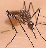
The bite of the female Aedes aegypti mosquito can transmit the virus that causes Dengue fever to humans
The bite of the female Aedes aegypti mosquito can transmit the virus that causes Dengue fever to humans.

The days of the flying mosquito may be drawing to a close.  In a study published in the February 22nd issue of the Proceedings of the National Academy of Science, a team of American and British researchers report that they have engineered a mosquito in the lab that produces offspring that cannot fly and consequently cannot infect humans with the virus that causes Dengue fever (full article).  Their findings may lead to a sustainable mosquito population suppression strategy that dramatically reduces human morbidity and mortality from a variety of diseases transmitted by mosquitoes.

In this study, the scientists specifically focused on the Aedes aegypti mosquito that causes Dengue fever.  The researchers manipulated the genetic material of the males of this species in the lab to carry a novel trait:  the inability to fly.  When these modified males were mated with normal, wild-type females, they passed the trait on to their female offspring.  By rendering the female offspring flightless, the scientists effectively imposed a death sentence on this group.  If the females cannot fly, they cannot elude predators, mate with males, escape from water, or seek out human blood.  Most importantly a flightless female may lead to the eradication of Dengue fever, since the disease is transmitted by the bite of female Aedes aegypti mosquitos.

The researchers predict that 6-9 months after introducing the modified males into the wild, the wild-type females in the area will be completely replaced by the flightless offspring of the modified males.  This is big news with important applications in the control of mosquito-borne disease.  This method of control offers several advantages over traditional techniques because it specifically targets the species of mosquito that causes Dengue fever and bypasses the use of toxic insecticides.  And as senior author Luke Alphey notes, “Another attractive feature of this method is that it’s egalitarian: all people in the treated areas are equally protected, regardless of their wealth, power or education.”

The next step for the researchers is to study the mating competitiveness of the modified males in the wild and whether their flightless female offspring will actually suppress the wild-type population as predicted.  Additionally, the methods used by these scientists could be adopted to control other species of mosquitoes that spread serious diseases such as West Nile virus and malaria.

Dengue fever is a flu-like illness with no vaccine or treatment that infects 50-100 million people each year in over 100 countries in tropical and subtropical climates, including Puerto Rico and tourist destinations in Latin America and Southeast Asia.  It is the most common mosquito-borne disease and the CDC estimates that one third of the world’s population lives in areas where the disease is endemic.  Other diseases transmitted by mosquitoes include West Nile virus, malaria, Rift Valley Fever, and Yellow fever.  Taken together, these illnesses represent growing public health issues that require effective and sustainable mosquito population control measures.  The flightless mosquito may just be the answer to this urgent problem.

Want more information?

Press release

CDC site on Dengue fever

Drugs That Kill Bacteria Can Also Make Them Stronger

By Johnathan Nieves ‘11

A recent study showed that exposure to low levels of antibiotics increased mutations in bacteria hundreds of times more than normal, making the creation of drug-resistant bacteria more likely. A drug under development by Radnor, PA-based PolyMedix, Inc. shows promise for addressing the serious threat of drug resistance by mimicking the human body’s defenses.

Click on the Image above to see Polymedix's Drug In Action (Sources: Image, World Health Organization; Video, Polymedix, Inc. )

If you don’t take your prescription antibiotics as your doctor advises, then listen up. Just last week (February 12) a paper published in the journal Molecular Cell described how exposure to low levels of antibiotics increased mutations in bacteria hundreds of times more than normal, making the creation of drug-resistant bacteria more likely. A drug currently under development by Radnor, PA-based PolyMedix, Inc., however, shows promise for addressing the serious threat of drug resistance by mimicking the human body’s defenses.

Drug resistance has been a growing health concern for decades now since the introduction of penicillin in the 1940s, the first available antibiotic of its kind. Drug resistance occurs because of bacteria’s natural ability to evolve through mutations it incurs as it reproduces. As it turns out, researchers have found that low antibiotic dosages are triggers for increasing the rate at which bacteria mutate, thus, increasing the likelihood of drug resistance.

“Like anything in nature, bacteria have ways to fight its opponents, and do so either by pumping antibiotics out of themselves through a process called efflux, or by rapidly mutating and changing the shape of the target of attack of the antibiotic drug. They can do this, even with large doses of antibiotics, it’s their innate way to try to survive,” explains Bozena Korczak, Vice President of Drug Development at PolyMedix Inc..”

“Upping the antibiotic dosage may be a viable solution but not the ultimate one,” adds Korczak. Driven by science conducted at the University of Pennsylvania, PolyMedix is investigating a new type of antibiotic drug that works by imitating the human immune system.

PolyMedix’s investigational antibiotic agent, called PMX-30063, is the first of its kind with a new approach to address the serious health implications of drug resistance by mimicking host defense protiens. Unlike most antibiotics, host defense proteins work fundamentally different. Rather than crossing the bacterial membrane to find a target like most antibiotics, they selectively target the cell membranes integrity by poking holes into it. This diminishes the bacteria’s ability to remain intact and the bacteria and its internal components become degraded (See video demonstration by clicking on the image above).

Polymedix purports that this unique mechanism of action makes drug resistance unlikely to develop. Korczak insists that “the best approach to preventing this phenomenon is by directly attacking the bacteria’s cell membrane, rendering them destroyed and dead in a way that there is little chance of resistance.”

To study the ability of bacteria to resist an antibiotic drug, a laboratory experimental method known as “serial passage” is used by intentionally trying to create bacterial drug resistance. Using this experiment, PolyMedix has shown that resistance did not appear to its compounds in contrast to traditional antibiotics.

So far, data from two Phase I clinical studies demonstrate that the compound is safe and well-tolerated. PolyMedix is on schedule to complete the third and final segment of the ongoing Phase 1 study with PMX-30063 early this year and commence Phase 2 studies later this year.

PolyMedix has received 9 grants and research contracts from the National Institutes of Health and branches of the military to help support the development of its antibiotic compounds.

To view the press release associated with this piece, please click here.

To learn more about PolyMedix, Inc., please visit www.polymedix.com.

Flu to the rescue!

By Shelly Hwang

February 17, 2010

It’s flu season, and with the H1N1 virus being the spotlight of current news and the CDC pushing for nation-wide flu vaccination, people have become terrified of the influenza virus. However, a recent study done by researchers at the Yale University School of Medicine (published in the February 18 issue of Cell Host and Microbe) revealed that the stress response caused by the flu actually protects against death by secondary infection by using mice with bacterial infections.

Influenza can damage the lungs but usually does not kill. However, secondary infections such as pneumonia can occur after infection with the influenza virus and are much more deadly. Each year, more than 200,000 U.S. residents are hospitalized for flu-related complications, and about 36,000 Americans die on average per year from complications of the flu (CDC Statistics).

While previous studies on the flu have shown repressing of the immune system, such studies have only studied a single pathogen and focused on local effects of influenza at the site of infection. In reality, organisms are exposed to multiple infectious agents at a time and the effect of influenza on the whole immune system has not been studied.

This study, led by Dr. Rusian Medzhitov from the Department of Immunology, used a mouse model to examine the effects of the lung infection caused by influenza on the immune response to bacterial infection. Surprisingly, the researchers found that the influenza lung infection led to increased production of glucocorticoids (GC), which are produced in response to stress and known to play a key role in regulating inflammation. They found that virus-induced GC production is essential to controlling inflammation, as shown by the death of mice lacking GC’s that were infected by multiple pathogens.

So the next time you find yourself miserable and overwhelmed with the unpleasant flu FACTS symptoms (Fever, Aches, Chills, Tiredness, Sudden symptoms), remember to thank the virus for protecting you from fatal secondary infections.

Press Release

Department of Immunobiology at Yale

The key to locking up cancer

Sara Braniecki

Structure of TGF-B

In the current issue of Nature Cell Biology, a team of researchers led by Philip Howe from the Department of Cancer Biology at the Lerner Research Institute explain how they worked backwards to discover the protein that triggers cancer cells to be released from the original tumor, thus giving rise to new tumors.  Knowing this can lead to the development of drugs that contain cancer to one location, making it more efficiently treatable.

The researchers already knew that a process called epithelial-mesenchymal transdifferentiation (EMT) was important for cells on the surface of a tumor to transform into cells that are able to grow a new tumor elsewhere in the body.  The researchers worked backwards through the EMT process to find out what initiates it.  The researchers discovered that a protein called disabled-2 (Dab2) activated the EMT process and Dab2’s formation was triggered by transforming growth factor-b (TGF-b).

The EMT process is often what leads to death in patients with breast, ovarian, pancreatic, and colon-rectal cancers.  With the information these researchers have discovered about cancer cells, researchers can now begin to create drugs to stop EMT and stop cancer from spreadings.  This information could also lead to understanding how other diseases progress and can be contained.

Further Information.

Bullet shaped virus may be the future of new cancer and HIV treatments

By Liz Humes

An obscure virus that does not harm human cells has been generating a wave of excitement in the scientific community.  So what is the big deal?  A team of researchers from UCLA has reported the 3D structure of the vesicular stomatitis virus (VSV) in a break-through study published in the February 5th edition of Science (full article).  Their findings may shed light on how VSV can be manipulated and used in the treatment of cancer and in the development of vaccines for HIV and other harmful viruses.

The researchers used advanced, cutting-edge imaging techniques to visualize the 3D structure of VSV, which appears to have a bullet shaped head and cylindrical trunk.  They also characterized how the virus comes to form this bullet shape.  With this additional level of understanding of the physical structure of the virus, scientists believe that they can find ways to modify the structure of the virus and use it to treat and prevent illnesses such as cancer and AIDS.

As author Z. Hong Zhou remarked, “This work moves our understanding of the biology of this large and medically important class of viruses ahead in a dramatic way.”

VSV is a model virus that scientists use in the laboratory to study dangerous viruses that cause illnesses such as the flu, measles, and rabies.  Previous studies have shown that VSV can detect and kill human cancer cells.  Other studies have addressed the question of how to manipulate the virus to deliver a vaccine against HIV to the human body.

3D animation of VSV trunk

(Video is a 3D animation of the lower trunk structure of VSV-source)

A closer look at vaccine technology

A current trend in vaccine development is to use harmless viruses as “vectors” that can carry a specific vaccine to human cells.  These viruses have been engineered in the laboratory to carry pieces of genetic material from other pathogens and when they attach to human cells, they inject this genetic material into the cells.  These actions mirror an infection by the pathogen itself, although the virus vector does not actually cause an infection, and stimulates an immune response.  The human body then remembers how to respond to this pathogen the next time it encounters the pathogen and the body is protected from infection.

Modified versions of the viruses that cause the common cold and small pox are being studied in addition to VSV for use as vaccine vectors.  Given the potential that this type of vaccination has to prevent deadly infections from viruses and bacteria, this is an area of research one should surely keep an eye on.

Want more information?

Press release

Vaccination information

HIV vaccination development