Alzheimer’s Afternoon Seminar Series
APP and Bioenergetics
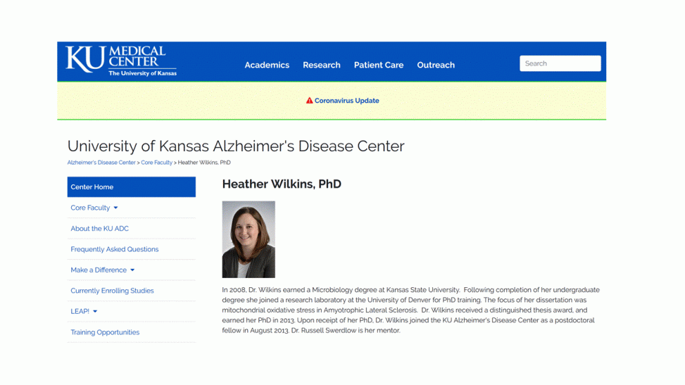
Dr. Heather Wilkins, University of Kansas Medical Center
April 23, 2020
Dr. Wilkins began by reviewing the “Amlyoid Cascade” and the “Phosphorylated Tau” theories of Alzheimer’s disease (AD), noting their similarities. She is, however, most interested in a third theory, called the “Mitochondrial Hypothesis” which posits that mitochondrial dysfunction is the first step in the development of Alzheimer’s Disease. She proposed that mitochondrial dysfunction eventually leads to the aggregation of AB and p-tau. While mitochondrial dysfunction is commonly caused by normal aging, Dr. Wilkins suggested that this occurs more rapidly in those predisposed to AD.
Today she focused on the association between mitochondrial dysfunction and the accumulation of Aβ.
She reviewed the processes by which the amyloid precursor protein (APP) is cleaved to form peptide fragments by the various secretase enzymes to generate Aβ chains of various length (with Aβ 40 and 42 being the most commonly studied). She also reviewed the “timeline of discovery” to note how our understanding of these processes has improved relatively recently.
These resources are available at: http://kualzheimer.org/
Dr. Wilkins explored the evidence that AD is primarily a problem with mitochondrial energy metabolism.
She noted, as support for this view, that AD pathology is associated with increases in mitochondrial DNA (mtDNA) mutations and deletions; a reduction in components of the electron transport chain, including cytochrome C oxidase (Complex IV); decreased glucose uptake and metabolism; and the presence of older, malfunctioning mitochondria.
Her work focuses specifically on the importance of the mitochondrial membrane potential.
A reminder for non-experts: mitochondria, often called the “powerhouse” of the cell produce ATP and other forms of useful stored energy by burning fuels such as sugars and fats.
Mitochondria act much like a battery, separating + and – electrical charges. This separation of charge occurs across the mitochondrial membrane. It separats two compartments – the internal space (the matrix; with a net negative charge) and external space (the intermembrane space, with a net positive charge). The burning (“oxidation”) of fuels provides the power to charge the “mitochondrial battery”. A fully charged mitochondria has an electrical potential of about 0.1 volts. When the two compartments are connected, the flow of protons (positively charged ions) powers the production of ATP.
In general, young mitochondria are superior for producing high amounts of ATP while minimizing the production of toxic byproducts, e.g. reactive oxygen species (ROS).
Dr. Wilkins proposed that the mitochondria membrane potential – specifically those that are too low (hypopolarization) or too high (hyperpolarization) – is linked to the amount of Aβ that accumulates in cells.
To test this, she studies SY5Y neuroblastoma cells, including mutant cell lines that lack key components of the mitochondrial “battery”. She also studies the impacts of drugs which change the polarization of mitochondrial membranes, such as FCCP which causes hypopolarization and oligomycin which causes hyperpolarization.
She observes that:
-
- Mutant cells lacking key proteins of the electron transport chain have reduced membrane polarization, reduced accumulations of Aβ 40 and 42, and lower beta-secretase levels.
- FCCP acts similarly to cause hypopolarization and reduce Aβ 40 and 42.
- Oligomycin treatments, expected to cause hyperpolarization, led to more Aβ 40 and 42.
In general Dr. Wilkins observes that the mitochondrial membrane potential is positively correlated with the accumulation of Aβ42.
She expanded this work to examine the response of neurons developed from stem cells (iPSCs) of patients confirmed to have had Alzheimer’s disease. The mitochondria of iPSCs from older AD patients did not exhibit altered mitochondrial membrane potentials but, interesting, those from younger patients did.
She then pivoted to discuss a theory called “MAGIC: Mitochondria As Guardians In the Cytoplasm”, described here:
Fang, E.F., Hou, Y., Palikaras, K. et al. Mitophagy inhibits amyloid-β and tau pathology and reverses cognitive deficits in models of Alzheimer’s disease. Nat Neurosci 22, 401–412 (2019). https://doi.org/10.1038/s41593-018-0332-9
This study showed that the recycling of old, poorly functioning mitochondria (“mitophagy”) is inhibited in AD. Those authors conclude: Our findings suggest that impaired removal of defective mitochondria is a pivotal event in AD pathogenesis and that mitophagy represents a potential therapeutic intervention.”
This theory emphasizes the importance of recycling old mitochondria to maintain efficient ATP production, without generating toxic byproducts. This strategy has been proposed to address a range of neurodegenerative disorders, as highlighted by a fun paper we sometimes discuss in class:
Komen JC, Thorburn DR. Turn up the power – pharmacological activation of mitochondrial biogenesis in mouse models. Br J Pharmacol. 2014;171(8):1818–1836. doi:10.1111/bph.12413
Or, if you want to more recent example, try: https://www.mdpi.com/2073-4409/9/1/150/htm#cite
Dr. Wilkins suggested that the APP protein, or parts of it, can clog mitochondrial membrane pores, causing dysfunction. She also noted that beta-secretase is required for the assembly of the mitochondrial electron transport chain.
She noted that mitophagy reduces the accumulation of Aβ 40 and 42, an interesting observation she hopes to explore using her neuronal iPSC system.
