Investigating the Anti-Inflammatory Effects of Japanese Traditional Medicine on T-cell-mediated Enteritis

Gillian Ferko and Katie Swade

Summary of:

Ueno N, Hasebe T, Kaneko A, Yamamoto M, Fujiya M, Kohgo Y, Kono T, Wang CZ, Yuan CS, Bissonnette M, Chang EB, Musch MW. TU-100 (Daikenchuto) and ginger ameliorate anti-CD3 antibody induced T cell-mediated murine enteritis: microbe-independent effects involving Akt and NF-κB suppression. PLoS One. 2014 May 23;9(5):e97456. doi: 10.1371/journal.pone.0097456.

Background

What is Enteritis?

Enteritis, more commonly known as Crohn’s disease, is a chronic inflammatory disorder of the digestive or GI tract that affects 1.6 million Americans. Crohn’s affects people of all ages, but the exact cause of the disease is unknown. It can affect anyone, but is usually diagnosed between ages of 15 and 35 for males and females equally. Possible causes could be genetic or environmental factors (antigens in the environment that can stimulate inflammation). Symptoms include frequent diarrhea, abdominal pain and cramping, rectal bleeding, fatigue, weight loss, reduced appetite, and fever. There are periods of high intense symptoms, but the disease does not persist constantly. Up to 20% of the people with Crohn’s have a blood relative with IBD (Inflammatory Bowel Disorder)

Screen Shot 2015-04-16 at 11.05.07 PM

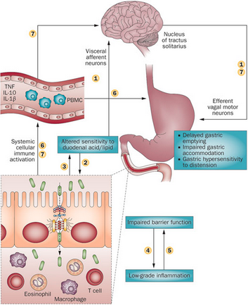
Figure 1. Schematic representation of the potential mechanism involved in the pathophysiology of Gastroenteritis. 

Enteritis can be induced in the lab by treating cells or mice with anti-CD3 antibodies.  These antibodies selectively activate T lymphocytes in the small intestine, which causes the pooling of intestinal contents, and eventually epithelial cell apoptosis. These antibodies have also been shown to increase TNF-⍺ levels in the small intestine and therefore are essential in the development of enteritis.  This anti-CD3 antibody response was first discovered in humans in an attempt to suppress organ transplant rejection.  Post-treatment, these patients developed a systemic cytokine response, indicating that this antibody leads to inflammation in the small intestine.

Japanese Traditional Medicine Background:

A traditional Japanese medicine, TU-100 (daikenchuto) is thought to have anti-inflammatory, pro-kinetic, and blood flow effects in the GI tract.  The three main components of TU-100 are ginseng, ginger, and Japanese green pepper so the complex is valued as an all-natural, environmental, and traditional form of medicine. Ginger has anti-inflammatory and blood flow effects that can potentially modulate mitogen activated protein kinase, protein kinase B (Akt), and NF-kB activities.


Question: Can TU-100 or its components block the effects of Anti-CD3 antibody-induced enteritis?

Approach: 

In all mouse studies, only germ-free (GF) and specific-pathogen free (SPF) mice were used to ensure the findings were independent of gut microbes.  Mice were given TU-100 or ginger alone by diet or gavage for up to 24 hours then treated with anti-CD3 antibodies.  The mice were then sacrificed and a laparotomy was performed to remove the intestine.  A segment of the intestine was removed, weighed, and stained.  RNA was extracted from this segment and analyzed for TNF-⍺ using Western blot and ELISA.

Additionally, human colonic adenocarcinoma (Caco2BBE) cells were treated with IFNy (100 U/ml) to increase TNF-⍺ receptor expression.  These cells were co-cultured and treated with TU-100 with Human Jurkat cells to study the interaction and signaling between T-lymphocytes and epithelial cells.


Main Findings:

-Anti-CD3 antibodies were shown to cause a type of enteritis that is dependent on T cells and regulated by lymphocytes.  This enteritis can be classified as gut microbe-independent and independent of intestinal bacteria.

-TU-100 and ginger alone, but not ginseng or Japanese pepper alone, blocked the anti-CD3 antibody-induced enteritis by significantly decreasing the amount of fluid accumulation within the jejunum and the stimulated expression of TNF-⍺.

-TU-100 also decreased the resulting apoptosis from anti-CD3 antibody treatment in jejunal epithelial cells.

-TU-100 and ginger were shown to block the activation of Akt after anti-CD3 antibody treatment, consistent with findings that ginger has been reported to block Akt activation.

-In the T-lymphocyte and epithelial cell co-culture, TU-100 and ginger show a block in anti-CD3 antibody induced activation of T-lymphocyte Akt and subsequent TNF-⍺ activation of epithelial NFkB.

-These findings demonstrate that TU-100 and ginger do in fact block the effects of anti-CD3 antibody induced enteritis.

Broader Context:

Enteritis is classified as a Inflammatory Bowel Disorder (IBD), along with a disorder called Ulcerative Colitis.  Enteritis is associated with inflammation in any part of the the GI tract, while Ulcerative Colitis is associated with inflammation only in the colon or large intestine. These disorders, along with other small bowel inflammatory diseases like viral enteritis or Ciliac’s disease, remain unclear in regards to cure or effective treatment.   Understanding the pathophysiology of enteritis and the effects of TU-100, ginger, or other substances can help extend the potential therapies for gastrointestinal disorders.


References:

https://www.crohnsandcolitis.com/crohns

Changes in gastrointestinal tract function and structure in functional dyspepsia doi:10.1038/nrgastro.2012.255

Investigating the Role of Angptl4 in Proteinuria associated with Nephrotic Syndrome caused by Minimal Change Disease

Mike Aimino & Lisa Freeman

Summary of:

Clement, L.C., Avila-Casado, C., Macé, C., Soria, E., Bakker, W.W., Kersten, S. & Chugh, S.S. (2011). Podocyte-secreted angiopoietin-like-4 mediates proteinuria in glucocorticoid-sensitive nephrotic syndrome. Nature Medicine, 17(1), 117-122. doi: 10.1038/nm.2261.

What is Nephrotic Syndrome?

Nephrotic syndrome is a condition that causes proteinuria, hypoalbuminemia, edema, hyperlipidemia, and lipiduria in individuals that have it. Proetinuria and lipiduria are when there are proteins and lipids in the urine, respectively. Hypoalbuminemia is when there is a low amount of albumin in the blood while hyperlipidemia is when there are high amounts of lipids in the blood. It can be caused by diabetic nephropathy, minimal change disease (MCD), focal and segmental glomerulosclerosis (FSGS), or membranous nephropathy. MCD is the primary cause of nephrotic syndrome in pre-adolescents, making up 85-95% of the cases. About 15 in 100,000 children have MCD with 2-7 new cases annually in 100,000 children. The prevalence of MCD is much lower in adults, making up only 10-15% of the cases. MCD is sensitive to glucocorticoid treatment, while the other diseases show a varied response, making it a good target to study.

Other complications of nephrotic syndrome include foot process effacement of podocytes. Normally, podocytes extend primary processes to the glomerular basement membrane (GBM) of the capillaries. Foot processes extend from the primary processes and lie on the GBM. Adjacent foot processes then interdigitate, which looks similar to locking your fingers together (pictured below). In individuals with necrotic syndrome, the foot processes disappear, making it look like the cell membrane is continuous. This leaves spaces between podocytes which allows proteins to leave and enter the urine.

(National Institute of Diabetes and Digestive Kidney Diseases)

All angioproietin-like proteins (Angptl) are glycoproteins that are sensitive to glucocorticoids. Angiopoietin-like proteins have been found to play a role in the development of hypertriglyceridemia and tumor metastasis. They have many different effects on cells depending on the part of the body in which they are found. Angptl4 is an inhibitor of lipoprotein lipase and has an effect on triglyceride levels in the blood. There has been no previous research to show that Angptl4 plays a role in proteinuria.


Question

What role, if any, does Angptl4 play in proteinuria associated with nephrotic syndrome?


Approach

The researchers employed a large arsenal of experiments and analysis techniques, so we will only give an overview of the experimental approach here. They started by evaluating four different nephropathy models, each model simulating a different disease that causes nephrotic syndrome. They identified the model that yielded the greatest increase in Angptl4 expression (puromycin nephrosis, or PAN), and used this model in later experimentation. They then studied a previously established Angptl4 transgenic mouse model and developed two new transgenic rat models. The NPHS2-Angptl4 model is characterized by upregulated Angptl4 in podocytes, while the aP2-Angptl4 is characterized by an upregulation of circulating Angptl4, secreted from adipose tissue. They evaluated Angptl4 expression and morphological changes for each model.

Although data for the aP2-Angptl4 is presented in supplemental materials, the remainder of the paper is focused on continuing experimentation with the NPHS2-Angptl4 model. With this model, they measured albuminuria in rats at varying ages. They then induced PAN, a model for minimal change disease (MCD), and measured albuminuria again. To analyze an additional variable affecting protein expression, they treated the rats with glucocorticoids after inducing PAN and measured resulting proteinuria and Angptl4 expression. Finally, the researchers conducted an in vitro study in two different cell lines of the sialylation of Angptl4 on its electrophoretic migration and the level of proteinuria occurring in the transgenic models.

A variety of analysis techniques were utilized throughout the experiments described above. Light microscopy and electron microscopy were used to analyze morphological changes at the glomerular and cellular levels, respectively. Immunohistochemistry with confocal microscopy and immunogold electron microscopy were used to localize and quantify Angptl4 expression. The researchers used SDS-PAGE to detect urinary protein and a combination of 2-D gel electrophoresis and western blotting to differentiate between forms of Angptl4.


Main Findings

A key result from these experiments was that increased expression of podocyte-secreted Angptl4 (NPHS2-Angptl4 transgenic model) caused increased proteinuria, similarly to proteinuria observed in rat models and human MCD patients. Angptl4 expression was also associated with morphological changes characteristic of MCD. More specifically, proteinuria was induced when the glomerular basement membrane (GBM) showed the presence of Angptl4, even though the podocytes did not yet show morphological changes. The researchers interpreted these results as an indication that Angptl4 causes a defect in the GBM that ultimately leads to proteinuria. They also found that Angptl4 decreased after glucocorticoid treatment. Finally, their results showed that sialylation of Angptl4 was associated with decreased proteinuria.


Broader Context

The results show that in the PAN model there is a 60-80 fold upregulation of glomerular Angptl4 expression in the PAN model. This is close to the 120 fold increase found in the NPHS2-Angptl4 heterozygous, male rats, meaning that it is a good model for studying nephrotic syndrome. This model is the first demonstration of the important role Angptl4 plays in proteinuria. Because the NPHS2-Angptl4 rats had reduced albuminuria when fed with ManNac and had an increase in the sialylation of glomerular Angptl4, it suggests that hyposialylation could be a mechanism by which Anglt4 overexpression causes proteinuria. Therefore, treatment with sialic acid precursors could be a potential therapy for individuals with some forms of nephrotic syndrome, particularly minimal change disease.


References

1. Mansur, A., Georgescu, F., Lew, S. (2015). Minimal-Change Disease. Medscape. http://emedicine.medscape.com/article/243348-overview#a0199

Exploring Ciliary Mechanosensation as a Means to Understanding Polycystic Kidney Disease

Summary of:

Low, S. H., Vasanth, S., Larson, C. H., Mukherjee, S., Sharma, N., Kinter, M. T., Kane, M. E., Obara, T. & Weimbs, T. (2006). Polycystin-1, STAT6, and P100 Function in a Pathway that Transduces Ciliary Mechanosensation and Is Activated in Polycystic Kidney Disease


By: Lizz Reese and JT Stoner


What is ADPKD?

Autosomal dominant polycystic kidney disease (ADPKD) is one of the most common forms of polycystic kidney disease. It is known to occur in individuals and families of all different races and is estimated to currently impact the lives of 400,000+ people in the US. ADPKD usually presents in adulthood with about 50% of patients developing end-stage kidney disease by the age of 60.

This disease is characterized by fluid-filled cyst growth in both kidneys, which begin to replace much of the normal kidney mass. As a result, reduced function and organ failure may occur, requiring the patient to undergo dialysis and/or kidney transplant. PKD can also cause cysts in the liver as well as additional problems in the heart and blood vessels of the brain.

While there is no definitive treatment for ADPKD, treatments have been tailored to mitigate  non-kidney symptoms (e.g. blood pressure) and pain (e.g. treated with painkillers and antidepressants). Kidney-related treatments typically aim to control buildup of acid and to prevent elevated phosphate levels.


What causes ADPKD?

ADPKD is genetically inherited in an  autosomal dominant fashion. The mutated gene (either PKD1 or PKD2) causes renal cells to proliferate abnormally, resulting in the formation of fluid-filled cysts which eventually replace most of the normal renal tissue and lead to renal failure.


What is known?

Mutations in PKD1 or PKD2 are recognized as the underlying causes of ADPKD, with PKD1 being mutated in 85% of the cases. The function of the gene product, PC1, is poorly understood.  PC1 is a large, integral membrane protein which is believed to have extracytoplasmic ligand binding domains, although ligands have yet to be identified.  The C-terminal tail has been implicated in signal transduction pathways such as the wnt pathway, a pathway leading to AP-1 transcription factor activation, G protein signaling, calcium signaling, and activation of STAT1. It is unknown, however, if any of these pathways are altered in ADPKD.

In a process that is thought to incorporate PC1, primary cilia of renal epithelial cells act as mechanosensors that respond to changes in lumenal fluid and flow (see figure below). Moreover, it has been shown in a number of case that defects in cilia proteins can lead to renal cystic diseases in humans and animals, though PC1’s role remains unclear.


Blog Post Picture V. Singla et al., Science (2006) Published by AAAS.


Article Summary

Autosomal dominant polycystic kidney disease (ADPKD) results from polycystin-1 (PC1) defects, which are poorly understood but known to implicate primary cilia. This paper identifies a novel mechanism of cilia function that leads to changes in gene expression via PC1 and shows that this pathway is inappropriately activated in ADPKD.  Under normal conditions, the PC1 cytoplasmic tail interacts with transcription factor STAT6 and the coactivator P100 to stimulate STAT6-dependent gene expression. Termination of apical fluid flow results in nuclear translocation of STAT6. Under ADPKD conditions cyst-lining cells exhibit higher levels of nuclear STAT6, P100, and the PC1 tail. Exogenous expression of human PC1 in zebrafish embryos results in cyst formation.


Hypothesis

The signaling pathway that transduces a mechanical signal from primary cilia to changes in gene expression is inappropriately activated in ADPKD by the proteolytic cleavage and nuclear translocation of polycystin-1 (PC1).


Methods & Models

A number of different cells lines and models were used throughout the experiment: MDCK renal epithelial cell line (derived from a canine), COS-7 cells (derived from monkey kidney tissue), and HEK293T cells (human embryonic kidney cells) as well as normal and diseased human and mouse kidneys and zebrafish embryos. The MDCK and COS-7 cells were transfected with FLS-PC1 or CTM-PC1 to demonstrate that the cytoplasmic tail of PC1 localizes to the nucleus and to show that the C-terminal half of the PC1 tail is cleaved, released from the membrane, and targeted to the nucleus. Results were viewed via Western blots and immunostaining. Additionally, MDCK cells were utilized to demonstrate that the PC1 tail interacts with P100 via CTM-PC1 or NTM-PC1 transfection, Coomassie staining, and confocal/immunofluorescence microscopy. MDKC and HEK293T cells were used in experiments to show that the C-terminal half of PC1 tail interacts with STAT6 and activates STAT6-dependent transcription by transfection with FLS-PC1 or luciferase reporter constructs, respectively, followed by Western blotting and reporter assays. MDCK cells continued to be used for experimentation to determine that STAT6 localized to cilia and translocated to the nucleus under ‘no-flow’ conditions via immunofluorescence microscopy with tagged STAT6. Immunohistochemistry was used to detect STAT6 in human ADPKD and normal kidneys. Primary cilia were detected with H&E staining. Zebrafish embryos were utilized to show that the human PC1 tail causes pronephric cysts by injecting one population with FLS-PC1 and monitoring human PC1 mRNA via RT-PCR at 3 days post fertilization.


Key Findings

Because the function of PC1 is poorly understood, these authors aimed to explore and better understand how its binding to transcription factor STAT6 and coactivator P100 affects the relationship between ciliary mechanosensation and the onset of autosomal dominant polycystic kidney disease.  The most significant discoveries in this study are outlined below:

  • Full length PC1 is vulnerable to rapid proteasomal degradation, localizes to the nucleoplasm, and is overexpressed in ADPKD conditions.
  • In a diseased kidney,  the C-terminal half of the PC1 tail is cleaved not only at a GPS domain, but another site as well, which leads to its release from the membrane.
  • Different constructs of PC1 localize to either the cytoplasm (NTS-PC1) or the nucleus (CTS-PC1 and CTSP-PC1), the latter of which are mediated by the C-terminal half of the PC1 tail, suggesting that the tail undergoes nuclear shuttling.
  • P100, which is a coactivator to transcription factor STAT6, binds to the PC1 tail. It  localizes to the basal body and primary cilia in polarized MDCK cells and is overexpressed in ADPKD renal tissue, specifically in cyst-lining epithelial cells.
  • In addition to PC1 binding with P100, it also binds with transcription factor STAT6 to stimulate STAT6-dependent transcription. More specifically, the C-terminal half of the cytoplasmic tail positively regulates STAT6-dependent transcription.
  • STAT6 was found to be moderately expressed in renal epithelial cells of normal human kidneys, overexpressed in cyst-lining epithelial cells of ADPKD kidneys, and is involved in transduction of mechanical signal originating at primary cilia. It is activated under no-flow conditions, translocating from primary cilia to nuclei of renal epithelial cells. Further experimentation suggests that STAT6-dependent gene expression is highly upregulated in ADPKD.
  • Using a zebrafish model, the authors were able to demonstrate that exogenous overexpression of soluble PC1 tail alone can stimulate renal cyst formation.


Broader Context

The primary findings outlined above have allowed the authors to construct the following signaling mechanism, which demonstrates how PC1 modulates STAT6-dependent transcription. Upon initial cleavage of the cytoplasmic tail of PC1,  the membrane-bound C-terminal half of the tail is freed from the membrane. It can then bind to STAT6 and the transcriptional coactivator P100 and translocate into the nucleus where it induces STAT6-dependent transcription. Evidence demonstrating that, in the absence of apical fluid flow, STAT6 translocates from primary cilia to the nucleus allowed the authors to hypothesize that the PC1/STAT6/P100 pathway may have greater implications in the relationship between mechanical signal transduction and transcriptional response. Moreover, because this pathway is highly upregulated in human ADPKD cysts, it is thought to have a significant role in the progression of this human disease. With a better understanding of how PC1/STAT6/P100 pathway is regulated, researchers can now focus their attention on specific components of this pathway that may serve as therapeutic targets for the treatment of ADPKD.


Links

ADPKD: https://www.genome.gov/20019622

Polycystins and mechanosensation: http://onlinelibrary.wiley.com/doi/10.1002/bies.20069/epdf


Degeneration and Disconnect: Restoring SMN Expression through SR Proteins in Spinal Muscular Atrophy

Heather Geist and Juliana Schneider

Summary of:
Wee, CD, Havens, MA, Jodelka, FM, Hastings, ML. (2014). Targeting SR Proteins Improves SMN Expression in Spinal Muscular Atrophy Cells. PLoS ONE, 9(12). e115205. doi:10.1371/journal.pone.0115205

Background:

What is SMA?
Spinal muscular atrophy (SMA) is an autosomal recessive genetic disease that is one of the most common inherited causes of pediatric mortality. SMA has occurs in 1 in 6000 live births and has a carrier frequency of 1 in 40. This disease affects the voluntary muscle movement controls of the nervous system. SMA is categorized as a degenerative neuromuscular disease, which involves the loss of  motor neurons in the spinal cord. This in turn prevents the muscle from receiving signals, leading to muscular atrophy over time. The most common form of SMA is called chromosome 5 SMA, which has great variability in age of onset. For this reason it is typically classified into types 1 through 4. Our main focus will be on Type I SMA patients. Type I is correlated with the lowest level of functioning patients. Onset of symptoms in Type I patients usually occurs at birth or during infancy, leading to the greatest impact on motor function. The most drastically affected muscles are typically those closest to the center of the body and because of early onset, these individuals have high mortality rates.

What is SMA Caused by?
SMA is caused by deletions or mutations in the Survival of Motor Neuron I gene (SMN1) located on chromosome 5. SMN proteins are essential for conventional motor neuron function. If a deletion or mutation of SMN1 occurs, an unstable form of the SMN protein results. The SMN2 gene, also located on chromosome 5, has the ability to produce SMN protein at smaller volumes (Figure 1). The main aberration between SMN1 and 2 is the single nucleotide in exon7, which is thought to be an exon splice enhancer. As a result of this single nucleotide difference, SMN2 cannot compensate for the loss of SMN1. The SMN1 gene is telomeric. It contains more than 4 genes, so it is susceptible to rearrangements and deletions. SMN2, however, is centromeric and is not as prone to rearrangement. Interestingly, increasing the inclusion of exon7 escalates the abundance of SMN protein. The SMN2 gene, also located on chromosome 5, produces SMN protein at smaller volumes. Inclusion of exon7 has been shown to have efficacy in animal models of SMA and early human clinical trials.

SMA gene 1 and 2 differenceFigure 1. Representative diagram of SMN1 and SMN2 genes and subsequent proteins in unaffected and SMA individuals. (Image modified from http://www.smasupportuk.org.uk/blog/ research/azzouz-laboratory-gene-therapy-for-sma).

Splicing and its role in SMA:
Exons and introns undergo a period of pre-mRNA splicing. This is conducted by the spliceosome complex, which is comprised of 5 snRNPs and other splicing proteins/factors (Figure 2). The splicesome complex works to identify exons and introns through binding to specific consensus splicing sequences on both the 5’ and 3’ ends of mRNA. Multiple proteins and sequence elements have been shown to play a role in regulating the splicing of SMN exon7. Specifically, SRSF1, SRSF2, and SRSF9 are known to influence exon7 inclusion, but few other members of the SR protein family have been explored regarding their role in SMN2 exon7 splicing. In addition to the SR protein family, the hnRNP proteins have also been implicated in play a role in exon 7 inclusion or exclusion.

Spliceosome review imageFigure 2. The spliceosome complex and process of pre-mRNA processing. (Image modified from http://www.nature.com/nrn/journal/v8/n11/box/nrn2237_BX1.html)

Question:
What characteristics and roles do SR and hnRNP proteins play in SMA disease pathogenesis? Do any of these proteins have therapeutic implications?

Experimental Approach:
Two main cell-lines were utilized throughout the experiments. The first was HeLa cells, which are derived from a cervical cancer tumor. The cells were treated and transfected with siRNA and Lipofectamine to knockdown specific SR and hnRNPs. In order to overexpress these proteins, expression vectors were transfected into the cells. The second cell-line used were human fibroblast cells derived from a Type I SMA patient only having one copy of SMN2. These cells were transfected as the HeLa cells were to knockdown the designated SR and hnRNP proteins. After cells were treated, RNA was isolated, and RT-PCR was conducted.

Proteins were extracted from the cells and immunoblots were performed by probing with 12 different antibodies directed at specific proteins. After quantitative analysis of fluorescence was performed, different levels of exon7 expression were observed. In vitro transcription was conducted on RNA splicing substrates with or without additional SR proteins. PCR analysis was done to quantify exon7 in these cells.

Key Results:
The most valuable result gathered from this experiment was the determined involvement of SRSF2 and SRSF3 in exon7 inclusion. Through knockdown of specific targets, an increased level of SMN protein was found to be present in these cells. This leads to further implications that these proteins could be therapeutic targets for Type I SMA patients.

Succeeding overexpression of SR proteins, the researchers observed a decrease in exon7 inclusion. Moreover, following successful knockdown of SR proteins in HeLa cells, it was found that exon7 inclusion increased significantly in 9 of 12 SR proteins. Specifically, exon7 inclusion was found to have the highest levels following SRSF3 knockdown. Following 40-50% knockdown of hnRNP A2B1 and U, a significant inclusion of exon7 resulted. This result confirmed that both SR and hnRNP proteins enhance splicing and through inhibiting their function, more exon 7 inclusion occurs.

Similar to the above experiments, the researchers decided to knockdown SR proteins in SMA cells and observe not only the effects on exon7 inclusion, but also SMN protein abundance. It was observed that upon knockdown of SRSF2 and 3, SMN protein levels increase. Thus, downregulation of these SR proteins leads to an increase in SMN protein levels. These results were consistent with the results obtained from HeLa cells. It was through this data that the researchers decided to examine the SRSF2 and 3 proteins more closely and deemed them to be the most effective therapeutic targets.

Broader Context:
There is a lower toxicity associated with targeting individual proteins such as splicing factors that have fundamental functions in the cell. Downregulation of single or multiple SR protein inhibitors of SMN2 exon7 inclusion could result in an improvement in full-length SMN protein. As a result, levels of SMN expression could increase to adequate therapeutic levels without fully disrupting other necessary functions of splicing factors.

Additional References:

MDA: Fighting Muscular Disease Website: mda.org/disease/spinal-muscular-atrophy/overview
Gene Cards: The Human Gene compendium Website: www.genecards.org

Therapeutic Benefits to SOD1 Silencing by way of AAV:miRNA Infection in ALS

Summary of:

Dirren, E., Aebischer, J., Rochat, C., Towne, C., Schneider, B. L., & Aebischer, P. (2015). SOD1 silencing in motoneurons or glia rescues neuromuscular function in ALS mice. Annals of Clinical and Translational Neurology. 

Brandon Goldson and Jessica Snyder

What is ALS?:

Also known as Lou Gehrig’s disease,  ALS (Amyotrophic lateral sclerosis ) is a fatal and incurable disorder primarily characterized by rapid progressive degeneration of motor neurons in the brain and spinal cord. This degeneration of the motor neurons leads to immobility, paralysis and eventually death 3-5yrs after diagnosis. About 5,000 people are diagnosed with an estimated 30,000 people living with ALS in the United States each year. Though ALS occurs in the world with no racial or socioeconomic boundaries, risk factors include age, sex and genetics. Regarding genetics, 10% of the people suffering from ALS display an inherited form of the disease. Within that population, 20% present a mutation in the gene encoding superoxide dismutase 1(SOD1).


What is the Known Pathophysiology of ALS and SOD1?:

Over 160 mutations of SOD1 have been found to cause ALS. These mutations cause an increase in toxicity and a degeneration of motor neurons in the brain and spinal cord.


What is the Hypothesis?

MiR Sod1 and MiR -b SOD1 effectively silence SOD1 (g93A) activity in mice expressing the human mutated gene. This silencing improves function and delays degeneration and symptoms associated with ALS in mice.


What is the Experimental Design

The effect of a miRNA designed to silence the SOD1 gene was tested both in cell culture and a mice model.  Both models contained a SOD1 gene mutation.  After the silencing effect was confirmed in vitro,  the researchers tested the efficacy of two treatments in SOD1 transgenic mice.  The first treatment was AAV6:miR SOD1 and the second was a combination of AAV6:miR SOD1 and AAV9:miR SOD1.  In all but the final experiment, the mice were given the treatment soon after birth.


The effect of the treatment in the mouse model was tested in the following ways:

  • examine whether or not the AAV is localized in correct cells (motor neurons and astrocytes are test in the spinal cord)
  • Observe SOD1 expression with and without the AAV miR treatment
  • motor function test of mice with & without treatment (swimming latency test and rotarod test)
  • test of proteins contained within muscle sample
  • staining of spinal tissue to see motor neurons status (intact or not)  as well as the viability of neuromuscular junctions
  • muscle fiber thickness to evaluate whether or not muscle atrophy was lessened
  • Finally, adult transgenic SOD1 mice were given the treatment and evaluated for viability, motor function and the state of neuronal cells in the spinal cord.

What are the Key Results?:

1: Suppressing of SOD1 in astrocytes and motor neurons : Figure 1

  • Quantification of human SOD1 levels relative to miR control confirms that human SOD1 expression is significantly reduced in cells overexpressing miR SOD1 and miR-b SOD1.

2: Delays Disease Progression and Improved Disease Outcome in the following ways


  • Increase in intact spinal motor neurons:  Figure 4
    • NMJ function was rescued to 90% as compared to control G93ASOD1 mic
  • Rescues Neuromuscular junctions: Figure 4
    • As compared to WT and PBS-injected G93ASOD1 mice, significant preservation of motoneuron integrity at all levels of the spinal cord was observed in AAV6:miR SOD1 and AAV6 + AAV9:miR SOD1 groups.
  • Increase in muscle fiber diameter (rescues muscle atrophy): Figure 5
    • Hematoxylin and eosin staining of gastrocnemius muscle sections displayed maintenance of muscle integrity with respect to AAV9:miR SOD1 mice compared to control transgenic mice.


What is the Broader context?

These results show promise for the treatment of familial ALS.  The maintenance of motor neurons and NMJ is key to attenuating disease symptoms and improving quality of life in ALS patients. Full or partial rescue of neuromuscular function following neonatal and adult ICV infection of AAV-miR SODI may establish the therapeutic silencing approach as an effective treatment for ALS. Furthermore,  this paper confirms that SOD1 silencing in specific cell types such as motor neurons and astrocytes is crucial to this therapeutic targeting. Additionally AAV based therapeutic targeting may be used to alter other genes related to ALS prevalence.


P.S.  Helpful YouTube Videos about the Nervous System Below!

Cell Senescence and Inflammation: A Link to Atherosclerosis

Summary of:

Ito, T. K., Yokoyama, M., Yoshida, Y., Nojima, A., Kassai, H., Oishi, K., & … Minamino, T. (2014). A crucial role for CDC42 in senescence-associated inflammation and atherosclerosis. Plos One, 9(7). doi:10.1371/journal.pone.0102186


Trevor Griesman and Alissa Meister

Atherosclerosis is a chronic inflammatory disease affecting medium-large arteries beginning at birth, with the progression depending on many factors. The main risk factors for atherosclerosis include hypertension, hyperlipidemia, diabetes mellitus and others such as age, sex, smoking, and sedentary lifestyle. Possible complications from atherosclerosis include coronary artery disease, cerebrovascular diseases, and peripheral artery disease. Although these complications are responsible for over half of the yearly world mortality, they often occur late in the progression of the disease with limited early diagnoses.

The first molecular event in atherosclerosis is endothelial dysfunction in the arteries as a result of injury or inflammation. This dysfunction takes the form of either cell senescence, apoptosis, or activation. This paper focuses on senescence, when cells stop replicating. Atheroscletotic plaques result from an accumulation of lipids and smooth muscle cell proliferation. In response, endothelial cells over-express adhesion molecules and increase the recruitment of inflammatory cells. These  inflammatory cells further release cytokines, causing a cytokine-mediated progression of atherosclerosis and LDL oxidation.  Plaques deteriorate the cell wall and cause thickening of the surrounding muscles. Accumulation of these plaques can limit the flow of oxygen and nutrients to the rest of the body, leading to serious consequences depending on where these plaques form. Plaques forming in the coronary arteries cause Coronary Artery Disease (CAD), limiting the blood flow to the heart and increasing the risk for blood clots. Similar diseases can develop if these plaques form in the periphery, carotid artery, etc. Symptoms of atherosclerosis also depend on where these plaques form, and may include chest pain, weakness, numbness in the periphery, headache, kidney disease, and many more.

CDC42 is a GTPase in the Rho family responsible for cell cycle regulation functions such as migration, endocytosis, morphology, and cell-cycle progression. Additionally, CDC42 regulates the organization and proliferation of the actin cytoskeleton. Previous studies have shown that CDC42 may act in the senescence and inflammation of cells. The CDC42 pathway has been implicated as a potential therapeutic target for inflammation reduction in aging individuals, however this has yet to be studied in an in vivo model. Ito et al. (2014) set out to study the link between CDC42 and inflammation in senescent cells, and connect this to a potential role of CDC42 in atherosclerosis. The researchers specifically investigated the relationship between CDC42 and the inflammatory NF-κB pathway.


EXPERIMENTAL METHODS

In order to create a model for senescence, the researchers cloned the genes for p16 and p21 (cyclin-dependent kinase inhibitors) into retroviral vectors, and infected cells. This resulted in the integration of the gene DNA into the DNA of the cells. In order to determine the effects of different CDC42 pathway components, they knocked down genes using siRNA. They then used RT-qPCR (as we did in class) to measure the mRNA levels of three genes: the cytokine CCL2, the endothelial-leukocyte molecule E-selectin (SELE), and Vascular Cell Adhesion Molecule 1 (VCAM1). Western blotting was used to quantify translated levels of these proteins. For some experiments, other proteins, such as a deactivated form of CDC42 were upregulated in cells by infection with an adenoviral vector which contained the DNA that coded for those proteins. To measure innate immune response levels, induced-senescent cells transfected with various siRNAs were treated with LPS or TNF-α, and levels of CCL2, SELE, and VCAM1 mRNA were measured by RT-qPCR.

The researchers also studied mouse and roundworm models. Mice were made into conditional knockouts using a Cre-Lox system, in which special sequences called lox sequences are inserted into the genome flanking the target gene. The protein Cre cuts at the lox regions. A Cre protein was used that is inactive until treated with a drug, in this case tamoxifen. When the researchers introduced tamoxifen into their Cre-Lox mice, they effectively knocked out the gene between the lox sequences. The researchers targeted either CDC42, Mdm2 (a negative regulator of p53), or some combination of those genes. They also used Apolipoprotein E knockout mice. In these mice, they investigated protein expression in tissue sections using immunostaining. They recorded mRNA levels using RT-qPCR, and measured atherosclerotic lesion areas using tissue section staining. Finally, the researchers studied C. elegans worms, specifically the nol-6 strain, which has high innate immune expression due to a p53 dependant pathway. The researchers measured levels of immunity-gene mRNA using RT-qPCR in siRNA treated worms, and tracked the survival of the worms over time.


KEY RESULTS

The researchers found that in senescence-induced cells, the NF-κB pathway upregulates pro-inflammatory gene expression (Figure 1). This was shown through the creation of senescent like cells by introducing pathway kinase inhibitors, which led to an increase in inflammatory cytokine and adhesion molecule expression. Furthermore siRNA directly targeting this pathway decreased the inflammatory cytokines. The researchers additionally found that CDC42 also regulates pro-inflammatory gene expression, by testing knockdowns of the CDC42 pathway and observing a decrease in inflammatory molecules (Figure 2). After these two findings surrounding NF-κB and CDC42, the researchers further found that CDC42 up-regulates pro-inflammatory cytokines by activating NF-κB. However, when cells were activated by either LPS or TNF-α, CDC42 knockdowns had had cytokine levels higher than NF-κB knockdowns, implying that NF-κB is downstream of CDC42, as it functions with other methods of activation (Figure 3).

The researchers then decided to test these finding using in vivo models, including several strains of mice and C. elegans. An increase in pro-senescence signalling in atherosclerotic plaques was found, linking atherosclerosis and inflammation (Figure 4). Additionally, CDC42 deletion was found to reduce aortic infiltration by macrophages in atherosclerosis. Overall, using these in vivo models, the researchers found that CDC42 is a mediator of chronic inflammation, which leads to endothelial senescence.


BROADER CONTEXT

In senescent cells, replication is halted as cells age to prevent the onset of cancer or other genetic malfunctions. Senescent cells secrete inflammatory cytokines, possibly as a way of signalling to immune cells that will destroy the senescent cell before it becomes cancerous. As we discussed in class, one of the ways that endothelial cells can respond to stress is to become senescent. This can lead to the formation of plaques, as lipids and then immune cells infiltrate the tunica intima. The researchers investigated CDC42 because of its involvement in inflammation following senescence. In the context of plaque formation, if the senescent endothelial cells did not secrete inflammatory cytokines, less immune cells would invade the plaque, slowing the progression of the plaque and possibly preventing the formation of a fibrous cap. However, clinical treatments knocking out CDC42 are very far off, as the inhibition of all senescent inflammatory signalling could result in the buildup of senescent cells, and there is no way to target CDC42 specific siRNA to plaque sites. Additionally, the experiments were done in cell models of senescence, but other factors may contribute to senescence in vivo. 


References

Mitrovska, S. (2009). Atherosclerosis : Understanding Pathogenesis and Challenge forTreatment. New York: Nova Biomedical Books.

What Is Atherosclerosis? (2014, August 4). Retrieved March 6, 2015, from http://www.nhlbi.nih.gov/health/health-topics/topics/atherosclerosis


Is miRNA-30d a good pharmological target for treatment of diabetes?

Summary of: Zhao, X., Mohan, R., Ozcan, S., and Tang, X. (2012). MicroRNA-30d Induces Insulin Transcription Factor MafA and Insulin Production by Targeting Mitogen-activated Protein 4 Kinase 4 (MAP4K4) in Pancreatic β-Cells. The Journal of Biological Chemistry. Vol. 287, No. 37, pp. 31155 – 31164.

(written by Emma Frair and Yeana Jang)

What is diabetes? Diabetes is a metabolic condition in which a person’s body is either unable to produce or properly use insulin. A small organ near the stomach called the pancreas is responsible for producing and secreting all the insulin in one’s body.

Why is insulin so important? So… what’s the big deal if we don’t have insulin or if it doesn’t function properly? Well, we all know that the cells in our body that need a source of energy to funciton properly. That energy comes from food, which a majority of is turned into glucose or sugar serving as the source of our cell’s energy. Insulin is responsible for conducting and getting the glucose into the cells. Without proper insulin function one’s cells are not able to maintain energy in the necessary places.

What are these different types of diabetes?

Type 1- Only 5-10% of those diagnosed with diabetes have Type 1 diabetes. The type 1 diabetic patient’s pancrease produce little to no insulin. TREATMENT- These patients will have to have a daily injection or permanent pump to deliver his or her insulin. The blood glucose levels will have to be checked at least 4 to 5 times a day.

Type 2- Type 2 can also be a result from the depletion of insulin secretion; however, the difference from type 1 is that even if the patient’s pancreas is producing insulin his or her body has become resistant to its effects. TREATMENT- Type 2 diabetes can be treated with healthier eating and daily exercise. While the body remains resistant to insulin patients must treat it the same way as type 1 diabetics.

What do high glucose levels in the blood actually do?  Diabetes can still lead to serious health problems including: heart disease, blindness, kidney failure, and loweer-extremity amputations. Diabetes is also the seventh leading cause of death in the United Sates.

How is this related to the study of the inflammatory system? The β-cells in the pancreas are responsible for exclusively expressing and secreting the insulin transcription factors, PDX-1 and MafA. The inflammatory cytokine, TNF-⍺, is also secreted from the β-cells in the pancreas.

How do microRNAs work? microRNAs are a class of small non-coding RNAs that regulate function of a gene by binding the untranslated (UTR) end of the target gene and inhibiting the translation or secretion of that gene. It has been found that the relative high levels of mRNA-375 is responsible for inhibiting the glucose-induced insulin secretion. High levels of miRNA-375 (and any levels of miRNA-21, 34A, and 146) lead to negative regulation of insulin transcription factors made in the β-cells of the pancreas, resulting in a decrease of insulin production.

This is where the story begins…

It was hypothesized that the upregulation of miRNA-30d results in insulin production, which protects any β-cell functions that are impaired by the TNF-α cytokines. Intentional overexpression of miR-30d is beneficial in the prevention of diabetes.

In order to test this hypothesis, an insulin-secreting cell line MIN6 was transfected with siRNA against MAP4K4 compared with the negative control. This was analyzed by real-time PCR after incubation of TNF-α. The protein and mRNA levels of insulin increased when the levels of miR-30d and MafA were also increased, but there was a reduction of both TNF-α and MAP4K4. The expression of MafA and IRS2 could effectively inhibit TNF-α by silencing MAP4K4 (Figure 6).

Furthermore, pancreas of 10-week-old diabetic mice and heterozygous (normal) mice were isolated to analyze by MAP4K4 antisense LNA probe. Western blog and real-time PCR were used to further analyze. The normal mice had increased expression of miR-30d whereas the diabetic mice had decreased expression, and it was vice versa with M4P4K4 (Figure 7).

Overall, it is important to understand the signaling pathway of insulin production in β-cells by observing the roles of miRNA and MafA. Studying this mechanism could potentially provide new therapeutic agents for diabetes.

Estrogen has a Protective Role in Influenza A Virus Infection

Summary of:

Robinson, D.P., Hall, O.J., Nilles, T.L., Bream, J.H., and Klein, S.L. (2014). 17β-estradiol Protects Females against Influenza by Recruiting Neutrophils and Increasing Virus-Specific CD8 T Cell Responses in the Lungs. Journal of Virology. Vol. 88:9 p. 4711-4720. http://jvi.asm.org/content/88/9/4711.long

Influenza viruses cause contagious respiratory illness with a sudden onset of high fever, cough, sore throat, runny or stuffy nose, body aches, headache, and fatigue. There are 3 types of influenza viruses (A, B, and C) with influenza A and B viruses causing seasonal epidemics of disease. The Centers for Disease Control (CDC) estimates that flu-associated deaths range from 3000-49,000 each year depending on length and severity of the season (see the CDC website for more general information about the flu virus, http://www.cdc.gov/flu/about/disease/us_flu-related_deaths.htm). Moreover, pulmonary inflammatory diseases such as influenza are known to differentially affect males and females with women suffering a worse outcome. Prior evidence indicates that estrogen impacts the function of various immune cells and therefore may contribute to the differential outcomes between men and women following respiratory infections. The authors of this paper aimed to examine the role of estradiol (E2, one of the 2 major biologically active estrogens in non-pregnant humans) in both the host response to the influenza A virus (IAV) and the ability of IAV to replicate in the host. It was hypothesized that estradiol alters the recruitment and activity of immune cells therefore affecting the outcome of IAV infection. In order to test this hypothesis, female C57BL/6 mice were ovariectomized and implanted with either E2 or placebo capsules followed by infection with IAV. E2 treatment resulted in less morbidity as compared to placebo-treated females. E2 did not affect virus replication, but increased the levels of chemokines in lung homogenates, increased recruitment of neutrophils to the lungs, and increased the levels of interferon-γ and tumor necrosis factor-α released from virus-specific CD8 T cells. These effects were dependent on neutrophils because depletion of neutrophils in females treated with E2 increased morbidity, reduced chemokine production, and reduced CD8 T cell IFN-g production. In summary, sustained E2 levels affect the host response to IAV infection through a neutrophil-dependent mechanism leading to improved outcomes. This work sets the stage for further mechanistic studies related to how E2 regulates these effects and supports the consideration of E2 levels in the treatment of IAV infected patients.

Understanding the Complex Role of Isoprenoid Depletion in MKD

Summary of:

Burgh, Pervolaraki, Turkenburg, Waterham, Frenkel, & Boes. Unprenylated RhoA Contributes to IL-1b Hypersecretion in Mevalonate Kinase Deficiency Model through Stimulation of Rac1 Activity. The Journal of Biological Chemistry Vol. 289, No. 40, pp. 27757-27765, October 2014.

Hereditary autoinflammatory disorders are caused by genetic mutations in molecules involved in regulating the innate immune response. They are characterized by recurrent and usually short attacks of joint pain, rashes, abdominal pain, and fever. One category of these diseases includes hyperimmunoglobulinemia D and periodic fever syndrome (HIDS) and mevalonic aciduria (MA) and is caused by mutations in mevalonate kinase, an enzyme that acts upstream in the mevalonate pathway (Figure 1). Mevalonate kinase is responsible for the production of both non-sterol (farnesylpyrophosphate (FPP) and geranylgeranylpyrophosphate (GGPP) and sterol isoprenoids (cholesterol). Taken together, these disorders are referred to as mevalonate kinase deficiency (MKD). Patients with MKD suffer from periodic fever episodes, which have been attributed, at least in part, to depletion GGPP and subsequent uncontrolled release of the cytokine interleukin (IL)-1b from monocytes and macrophages.

Figure 1 (Clin Cancer Res, July 1, 2012 vol. 18 no. 13 3524-3531)


How does GGPP control IL-1b release? GGPP is a lipid moiety that is responsible for the prenylation of proteins including the Rho and Rab families of small GTPases, which cycle between a GTP bound active state and a GDP bound inactive state. Modification of these proteins by GGPP is involved in subcellular localization (membrane attachment) and the control of GTP hydrolysis via interaction with guanine nucleotide exchange factors (GEFs), GTPase activating proteins (GAPs), and GDP dissociation inhibitors (GDIs). GEFs facilitate the exchange of GDP for GTP to generate the activated form. GAPs increase the GTPase activity thereby causing inactivation. Finally, GDIs interact with the prenylated, GDP bound form to inhibit GEF/GAP binding and membrane localization (Figure 2). Lack of prenylation by GGPP has the potential to either activate (for example by blocking GDI interaction) or inhibit (for example by disrupting proper localization) GTPases depending on the circumstances.


Figure 2 (Genes & Dev. 2002, 16: 1587-1609)

In MKD cell culture models, Rac1, a member of the Rho family of GTPases, is known to have reduced prenylation by GGPP leading to increased activity, which has previously been reported to mediate IL-1b hypersecretion. However, activation of Rac1 does not account for all MKD related phenotypes and the exact mechanism of RacI activation has not been determined. This article explores the role of protein prenylation in the function of another Rho family member, RhoA, and hypothesizes that lack of RhoA prenylation contributes to some MKD phenotypes. In order to test this hypothesis, a cell culture model of MKD was used where THP-1 human moncytic cells were treated with simvastatin, a drug that inhibits the enzyme directly upstream of mevalonate kinase, HMG-CoA reductase. As previously observed, simvastatin treatment resulted in increased GTP bound Rac1, indicative of activation. However, simvastatin treatment resulted in decreased GTP-RhoA, indicative of decreased activation. In addition, inhibition of RhoA with C3 transferase (an inhibitor more specific for RhoA than simvastatin) resulted in increased GTP-bound (and therefore activated) Rac1 and IL-1b release. Taken together, these results indicate that decreased prenylation of RhoA leads to inactivation, which subsequently leads to increased Rac1 activation and IL-1b release. Ultimately, this study increased our understanding of molecular mechanisms related to MKD pathogenesis by adding another piece to the puzzle related to IL-1b hypersecretion therefore uncovering a novel target for treatment.