The rising cost of poor diet: ADHD symptoms in children

A handful of Halloween candy later, and you are skipping around the room, your hands are fidgety, speech is jittery, and it’s hard to contain your burst of energy. You are probably quite familiar with the sugar rush that affects you this one day a year… two days a year? Three days? Almost every day?

Time and again, nutritional studies and long-term research has shown that poor dietary patterns of children influence neural development and behavior, especially hyperactivity and attention capacities, leading to diagnoses like ADHD.

A study from 2009 followed a group of children over several years and found that for significant increases in “junk food” consumption at ages 4-5, the risk for hyperactivity at age 7 was elevated. Another study, from 2004, demonstrated a prolonged association between artificial food coloring and hyperactivity. While the cause of ADHD is still undetermined, dietary patterns certainly play a role. Not only do high-fat, high-sugar diets cause issues in the developing nervous system, but these diets tend to also be low in valuable vitamins and micronutrients.

A more recent study, published online in March 2018 in European Journal of Clinical Nutrition, showed a positive correlation between a “processed” food pattern, a “snack” food pattern, and ADHD symptoms in children age 3-6. On the other hand, there was a negative correlation between a “vegetarian” food pattern and ADHD symptoms. This study used data collected from over 14,000 preschoolers in Ma’anshan City, China, and was the first large scale study in mainland China to investigate connections between diet and ADHD in children. The prevalence of ADHD symptoms in the group studied was 8.6%.

Researchers asked caregivers and parents to answer questionnaires about the recent food consumption and food choices of their children, and gave the children a Conners Abbreviated Symptom Questionnaire to assess for ADHD symptoms. Then, the researchers allocated five dietary patterns to represent the common answers expressed by the caregivers and parents: (1) “processed,” for fast food, fried foods, preserved fruit, and other high-fat food items, (2) “snack,” for high-sugar foods like sweets, biscuits, cakes, and chocolates, (3) “protein,” for red meat, poultry, eggs, fish, fruit, and rice, (4) “beverage,” for flavored milk, soda, and yogurt, and (5) “vegetarian,” for grains, beans, and fruit or vegetable juice.

Children who had a “processed” or “snack” dietary pattern had a significantly higher likelihood of demonstrating ADHD symptoms, hyperactivity, and attention problems. There was no correlation between ADHD symptoms and the “protein” or “beverage” categories, but the “vegetarian” dietary pattern seems to act as a protector against ADHD symptoms.

This study can not show causation, or that a poor diet causes ADHD. However, it can not be refuted that diet is an influencing factor in development and behavior in children. High-fat, high-sugar foods tend to be cheaper, more accessible, and more conveniently packaged in bags and wrappers for on-the-go, and they don’t need to be refrigerated. They taste good. But, the cost is a rising prevalence of ADHD and similar disorders. The relationship between food and hyperactivity is further evident through studies that have shown that elimination diets and fish oil supplements can reverse the ADHD symptoms. Fatty foods and sugary foods should be eaten in moderation; Halloween should not be occurring every day.

Source:

Yan, S., Cao, H., Gu, C., Ni, L., Tao, H., Shao, T., Xu, Y., & Tao, F. (2018). Dietary patterns are associated with attention-deficit/hyperactivity disorder (ADHD) symptoms among preschoolers in mainland China. European Journal of Clinical Nutrition [Published online March 13, 2018]. doi:10.1038/s41430-018-0131-0.

Tumors, No Longer a Guessing Game

What makes one tumor different from another? The word ‘cancer’ can be used to describe hundreds of maladies, from a misshapen mole, to sickle shaped blood cells, to massive tumors in the heart, lungs, or anywhere else. But what makes a heart tumor different from a lung tumor? And why are there four different types of lung cancer? The answer lies in the DNA of each cancer cell.

Currently, doctors diagnose different types of cancer based primarily on where they are, what they look like in scans, and what molecules they are made of when samples are taken in biopsies. A new study introduces a novel method, looking at the methylation of cancer DNA, a process that inhibits or expresses specific functions in a cell that can characterize different disorders. By identifying the specific DNA methylations in each tumor, they can be attributed to a specific diagnosis; this is done simply by removing a small piece of the tumor, and analyzing it in the lab.

This study looked specifically at tumors on the central nervous system (CNS) and performed the methylated cancer DNA diagnostic test for 1104 patients, each diagnosed with one of 64 distinct cancers resulting in CNS tumors. In 76% of cancers, the test matched the existing diagnosis. In 12% of cases, the original diagnosis was revised as a result of this test. In the remaining instances, the test could not match to a known methylation class.

These results are extremely encouraging, and point to the end of subjective cancer diagnoses, where doctors must act as detectives, gathering clues to point to the most likely diagnosis. But this new test is a fingerprint; every cancer has a different one, and once they can be told apart, they need only consult a database stocked with thousands of cancer prints.

And this database is already live and free the world over, the data for every cancer case tested in this manner will be available and shared. Doctors will be able to readily compare the DNA in the cancers they encounter with other cases, which will likely lead to the discovery of new, unique cancers, and dramatically improve the accuracy of diagnoses overall.


Source:

Capper D, Jones DTW, Sill M, Hovestadt V, Schrimpf D, et al., (2018) DNA methylation-based classification of central nervous system tumours. Nature. doi:10.1038/nature26000

Immunotherapy a Potential Treatment for Breast Cancer

Breast Cancer Awareness

Immunotherapy is the treatment of a disease by changing the immune system. It was believed in the past that breast cancer did not trigger an immune response. Immune response is the reaction of cells of the immune system against a foreign substance that is not recognized as a part of the body. In early 2018, researchers at the University of Washington in Seattle published a study assessing the benefits and potential future of immunotherapy as a form of treatment for breast cancer. 

In the last decade, after the detection of an immune response in breast cancer patients, there have been numerous studies that have considered immunotherapy a possible treatment for this type of cancer. A research conducted on women 6 months prior to a breast cancer diagnosis showed high rates of T-cells (a type of white blood cell) against tumor associated proteins in women who would go on to develop breast cancer. Immune checkpoint inhibitor therapy is a mechanism that uses drugs to block certain proteins which prohibit immune cells from killing cancer cells. These agents allow T-cells to recognize cancer cells, and limit tumor growth by dividing and growing themselves. 

Research studies have shown that with the progression of cancer, the development of antigens (substances that trigger an immune response) diminishes, reducing immune response. This implied that in order to come up with a proper immunotherapeutic mechanism, researchers must focus on developing strategies that reverse this effect and increase immune response that promotes tumor destruction. One of the first breast cancer associated antigens was the MUC-1 (Human Mucin-1) protein. T-cells related to this protein were in low number in patients with the disease. So, boosting these numbers would be a potential therapeutic mechanism.

It should be noted that despite recent discoveries, breast cancer is still a poor producer of immune response. Tumor infiltrating lymphocytes (TIL), another type of white blood cells infiltrate tumor tissues and cause direct physical contact between them, which results in the physical destruction of tumor cells. However, its occurrence and concentration varies based on the type of breast cancer.

Studies are currently ongoing to identify factors that identify the patients who are most likely to benefit from immune checkpoint inhibitor therapies. This is aided by the fact that despite fluctuating amounts, TILs are always present to some extent. So, one strategy for successful patient identification is to increase the number of TIL beforehand. Immunization has been found as a possible way of accomplishing this. Research has shown that vaccine induced T-cells can travel to the tumor and induce an increase in TIL. Currently, there are several vaccines under development to target multiple breast cancer antigens at the same time to make treatment more effective.

In addition to immunization, standard therapies like radiation and chemotherapy can also increase the amount of TIL. As we understand more immune response inducing effects of traditional treatment methods, we can use them more effectively. Thus, the immune environment of the tumors can be used to combine standard and novel therapeutic strategies to develop more effective treatments. Research on such methodologies is still a field in progress and there is much to learn before we can use the knowledge of immune response in breast cancer cells to improve treatment approaches.

Reference: Disis, M.L., Stanton, S.E. 2018. Immunotherapy in Breast Cancer: An Introduction. The Breast 37:196-199.

Link to article

Link to image

Asthmatics Breathe a Sigh… of Exasperation

Asthmatics Breathe a Sigh… of Exasperation

Glucocorticoids are a class of steroids patients with asthma use the world over by way of an inhaler. They are one of several drug classes that can reliably save a persons life in the event of an asthmatic exacerbation. But as many know all too well, severe asthmatic episodes can still persist despite the application of this drug. Conventional wisdom among parents and doctors has been to increase the dosage of administered glucocorticoids in patients with more serious symptoms.

But a new study on asthmatic children across the United States by the National Heart, Blood, and Lung institute finds that there is no significant differences in severity of asthma symptoms or frequency of episodes across average and quintuple doses of glucocorticoids. Further, the study even suggests high doses may stunt growth in children, revealing the possibility of more serious problems in childhood use. The article was published in The New England of Medicine.

The year long study consisted of over 250 asthma sufferers aged 5 to 11 years old, and divided them into 2 groups of continued low-level use, and quintupled use. The experiment was carried out such that neither the patients, their parents, nor the doctors knew which treatment they were receiving.

The results suggest no significant difference between the occurrence of asthmatic exacerbations or their intensity regardless of how much glucocorticoid is administered. The study also collected data on the childrens height and weight during the period and found that the patients taking the quintuple dose displayed slightly shorter stature when controlling for various differences. This may be an indicator of serious developmental problems caused by continuous high use of the drug, and should be a concern for anyone taking or prescribing it.

This study highlights a need for more effective medications in asthma sufferers, and a better understanding of how existing medications work. Glucocorticoids are self-administered in the form of an inhaler, often on an as needed basis. Add to this the fact that children are the most heavily afflicted by asthma, and it becomes extremely easy for a patient to take too much of their prescription. Granted, quintuple dose is a far cry from the average, but when double or triple dose does not lessen the symptoms, one can tend to take too much. In an age of ultra targeted and molecular approaches to medicine, a more precise asthma treatments should be available, given the low threshold of effectiveness on glucocorticoids, let alone its possible ill affects.


Jackson DJ, Bacharier LB, Mauger DT, Boehmer S, Beigelman A, et al., (2018) Quintupling Inhaled Glucocorticoids to Prevent Childhood Asthma Exacerbations. DOI: 10.1056/NEJMoa1710988

What Would You Call a Reliable Tuberculosis Test?

TB testing machine

Latent Tuberculosis infection (LTI) is when an individual’s immune system shows a response to the bacteria that causes Tuberculosis, but the person has no clinical signs and is not infectious to others. If untreated, LTI has the chance of progressing into active tuberculosis. Tuberculosis Skin Test (TST) and blood test (IGRA) are the methods of detecting LTI. However, the results of these tests show vast differences, especially in patients with compromised immunity who have received the BCG vaccine against TB.

TST is conducted by putting a small amount of TB protein under the top skin layer of the inner forearm. Individuals with LTI show a firm red bump ≥ 5 mm when inspected after 48-72 hours. Researchers at Anakara University School of Medicine in Turkey conducted a study from 2013 to 2017 to check if higher cut-off values for TST (diameter of the red bump) would increase the specificity and agreement between the two tests. The study was conducted on three groups: all participants, solid organ transplantation (SOT) candidates, and patients schedules for anti-TNFα treatment (for people with immunosuppressive conditions like rheumatoid arthritis). All the subjects were BCG vaccinated. Patients with a history of active TB were excluded.

In order to test if a change in the cut-off value for TST would give better results, both TST and IGRA were conducted for all three groups at three different cutoff values. The diagnostic agreement was very poor for 5 mm and 15 mm but increased slightly for 10 mm in the anti-TNFα group. Overall, the results showed that although false positive results decreased with higher cut-off rates, false negative results increased.

TST is known to give false-positive results in BCG vaccinated individuals. 

Studies have shown that the type and timing of vaccination affect the TST response. In the US, BCG vaccines are given once during infancy and once during school age. Vaccination at infancy is believed to stop affecting TST after 10-15 years, however, repeated vaccinations after infancy are known to have a longer effect. Most of the differences in the two tests were observed to be positive for the skin test (TST) and negative for the blood test (IGRA). Researchers were of the opinion that the vaccines after infancy might have been the cause of this discrepancy. It was concluded that higher cut-off values for TST were not very effective in decreasing the variation between the two tests.

Analysis of the direct costs and probable consequences of the tests have shown a greater percentage of TST patients receiving preventive treatments due to the high possibility of false-positive results. This also means an increased waste of resources for unnecessary treatment. Moreover, there is the potential risk of numerous treatments leading to a resistance to TB-drugs. The rates are lower for IGRA alone and for two step screening strategies including both TST and IGRA. The American Thoracic Society (ATS) has stated that there is a lack of sufficient data to recommend any of these three methods, but specific guidelines for immunocompromised patients recommend IGRAs.

There is a need for more conclusive results to assess the reliability of the tests for LTI, especially in light of the inconvenience that TSTs cause for BCG vaccinated individuals.

Reference: Erol, S., Ciftci, F. A., Ciledag, A., Kaya, A., Kumbasar, O. O. 2018. Do higher cut-off values for tuberculin skin test increase the specificity and diagnostic agreement with interferon gamma release assays in immunocompromised Bacillus Calmette-Guerin vaccinated patients? Advances in Medical Sciences 63(2): 237-241

Link to article

Image

Will therapy benefit OCD patients? Computers have the answer.

fMRI machine
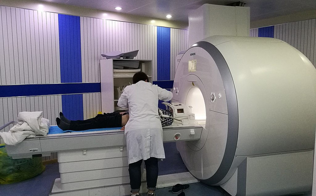
Patient being prepared for an fMRI. Credit: Ptrump16, Creative Commons.

Obsessive-compulsive disorder, or OCD, is characterized by unwanted, repetitive thoughts and impulsive, ritualistic actions. For example, a common fear among those with OCD is a fear of germs, which results in repetitive hand-washing. While historically OCD has been difficult to treat effectively, in recent years, modifications to cognitive-behavioral therapy have had more success. Cognitive-behavioral therapy is comprised of a series of sessions between a therapist and patient to identify negative thought patterns and symptoms, and address them through discussion, exposure to stress-inducing stimuli, and practice utilizing alternative coping mechanisms to ameliorate anxiety.

While cognitive-behavioral therapy can be effective, it is time-consuming and does not work for everyone. Using functional magnetic resonance imaging (fMRIs), scientists at UCLA trained a computer analysis system to study the brains of individuals with OCD, and determine which individuals were most likely to benefit from cognitive-behavioral therapy. Their study demonstrated that if an OCD patient were to receive a seven-minute fMRI scan, the computer program could predict the success of cognitive-behavioral therapy for that particular patient, at 67-70% accuracy.

For their study, researchers recruited 42 adults with OCD. All of the participants underwent fMRIs at the beginning of the study. Then, half of the participants attended cognitive-behavioral therapy sessions lasting about 90 minutes per session, five days a week for four weeks. At the end, their brains were analyzed with an fMRI again to detect any differences in structure or brain function. The other half of the participants were put on a four-week waitlist. At the end of four weeks, having received no therapy, their brains were scanned to see if there were any differences simply due to time. These participants received cognitive-behavioral therapy treatment after the four-week waiting period.

On the fMRI scans, the researchers were especially interested in studying the regions of the brain and their cellular networks that regulate attention, vision, motor skills, memory, self-evaluation, and the abstract sense of “mind-wandering,” or daydreaming, each of which play a role in development of OCD. They utilized mathematical models and computer learning to map differences between the participant’s brains, and match those results with behavior results of cognitive-behavioral therapy. They found that the computer could suggest which patients would benefit from therapy, regardless of individual symptoms or severity of symptoms.

fMRI brain scan
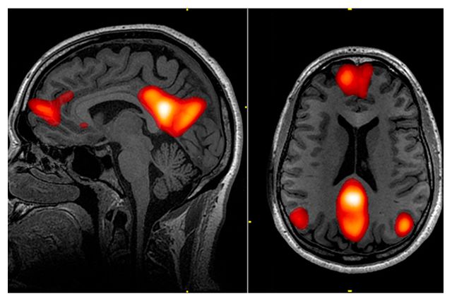
One of the brain networks studied was the default mode network, or DMN, which plays a role in “mind-wandering,” daydreaming, and abstract thought involved in thinking about the self. Regions of the DMN are highlighted in red in this fMRI scan. Credit: Leigh Hopper, UCLA Newsroom.

Widespread use of this predictive method would give therapists more information when deciding the best route of treatment for their patients. In the study, the researchers advocate for this fMRI computer model as a way to allocate time and resources, and direct cognitive-behavior therapy towards patients who are most likely to have success, versus other types of treatment such as medications, inpatient programs, intensive day programs, or group therapy. It is a move towards personalized medicine.

However, more research needs to be done to further advance this technique. Computers alone are not yet adequate to diagnose psychological disorders or comprehend subjective human experience. Furthermore, fMRIs are extremely expensive, and the money going towards fMRI scans could instead be put towards treatment. There is also a risk that those who the computer does not deem fit for cognitive-behavioral therapy miss out on a treatment opportunity that could actually help. While studies like this one advance scientific understanding of disorders like OCD, clinicians should proceed with caution when incorporating new computer-based evaluations that could be wrong and depersonalize the treatment experience.

Sources:

Reggente, N., Moody, T.D., Morfini, F., Sheen, C., Rissman, J., O’Neill, J., & Feusner, J.D. (2018) Multivariate resting-state functional connectivity predicts response to cognitive behavioral therapy in obsessive-compulsive disorder. PNAS [published online ahead of print]. https://doi.org/10.1073/pnas.1716686115.

Hopper, Leigh. 2018. Brain scan and artificial intelligence could help predict whether OCD will improve with treatment. UCLA Newsroom. Retrieved Feb. 5 from http://newsroom.ucla.edu/releases/brain-scan-AI-help-predict-ocd-improve-treatment.

Liquid Biopsy, Detecting Cancer DNA Before Tumors

A new blood test is capable of identifying genetic markers for eight major types of cancer. Though it only correctly identified cancer 70% of the time, this so-called “liquid biopsy” is a minimally invasive way to screen for cancer when no symptoms are present. It is at this stage cancer is at its most treatable, and at $500 costs no more than a colonoscopy, mammogram, or other cancer screenings.


The importance of such a test cannot be overstated in the fight against cancer. Insidious cancer cells can essentially lie dormant for 20-30 years before they grow into large, spread out masses that are difficult or impossible to eradicate. By identifying the cancer in its nascent stages, it can be usually be readily cured using radiation, surgery, or other common treatments. The difficulty lies in identifying the cancer DNA in patients blood, which is often present in miniscule amounts, and even when identified it can be difficult to trace the tissue of origin.

The eight major cancer types being screened here account for 60%, or 360,000, of all cancer deaths in the US last year, and there is nothing more critical to their effective treatment than an early diagnosis. Though as you can see from the accompanying chart, detectability varies; but this methodology is a work in process with large-scale human trials set to begin soon.

The research comes out of Johns Hopkins University in Baltimore and has been published in the journal Science. As this research begins large-scale human trials it will be evaluated for its utility in hospitals and doctors offices. The hope is that the principles underlying this methodology can be expanded to identify more types of cancer, and with greater accuracy. We may never be able to cure late stage cancer, and the treatments are often ghastly; but if the disease can be identified before it even forms tumors, the cancer can very likely be cured.


Cohen JD, Li L, Wang Y, Thoburn C, Afsari B, et. al. (2018) Detection and localization of surgically resectable cancers with a multi-analyte blood test. Science. 359(6378): 926-930


Molecular markers identified for autism, schizophrenia, and depression

Some psychological disorders, such as schizophrenia, tend to be highly heritable, meaning that the disorder is often passed down generationally within a family. Schizophrenia, for instance, is 60-87% heritable; if you were to have schizophrenia, there’s a 60-87% chance that one of your immediate relatives will develop symptoms, too. Similarly, major depressive disorder is 30-40% heritable. Therefore, in order to treat these disorders, its necessary to look at the genes involved. A February 2018 study published in Science found that there is significant overlap in gene expression between autism spectrum disorder, schizophrenia, and bipolar disorder, as well as an overlap between schizophrenia, bipolar disorder, and major depression. The strongest relationship was between schizophrenia and autism spectrum disorder.

Consider gene expression as a construction company. A construction company has a stockpile of materials: concrete, glass, cement, wood, nails, etc. The company has a crew of workers, and the crew is capable of building a variety of houses and apartment buildings. The construction company is analogous to the use of DNA by cells in the brain. The DNA is like the stockpile of materials. The materials are required to build anything, but the possible combinations of materials are endless. The RNA transcription mechanism in the cells is like the crew. The crew chooses which materials to use, and determines how much of each item is necessary for the project. In cells, this system is called “gene expression.” Every cell in the brain has the same DNA, or the same starting materials, but each cell has a different construction crew that decides to use the materials slightly differently; some build houses, some build apartment buildings, some build garages.

Instead of examining the DNA, or the building materials in over 700 cadaver brain samples used in the study, the researchers looked at the gene products, or what the construction crews built. It is unknown whether the gene products found in the brains caused the disorder symptoms, or gradually developed throughout life as the consequence of the disorders. But the study provides useful information regarding what proteins and structural factors manifest in disordered brains, and this information can be used to trace back to an origin point. Director of the UCLA Center for Autism Research and Treatment, and author of the study Daniel Geschwind said, “These findings provide a molecular, pathological signature of these disorders, which is a large step forward.”

The scientists found biological markers that tend to distinguish a brain with autism, for example, from the average brain. In the case of autism spectrum disorder, the study reported an increased activation of the CD11 gene, while another gene called CD2 was especially active in the brains suffering from depression. Additionally, the study mapped gene expression commonalities between brains with the same disorder, essentially establishing a molecular blueprint that can be recognized for diagnosis, and treated more effectively at the molecular level.

Sources:

Gandal, M.J., Haney, J.R., Neelroop, N.P., Leppa, V., Ramaswami, G., Hartl, C., Schork, A.J., Appadurai, V., Buil, A., Werge, T.M., Liu, C., White, K.P., CommonMind Consortium, PsychENCODE Consortium, iPSYCH-BOARD Working Group, Horvath, S., & Gerchwind, D.H. 2018. Shared molecular neuropathology across major psychiatric disorders parallels polygenic overlap. Science 359: 693–697.

Hopper, Leigh. 2018. Autism, schizophrenia, bipolar disorder share molecular traits, study finds. UCLA Newsroom. Retrieved Feb. 26 from http://newsroom.ucla.edu/releases/autism-schizophrenia-bipolar-disorder-share-molecular-traits-study-finds.

Seidel, D.C., Bulk, C., Stanley, M.A. 2017. Abnormal Psychology: A Scientist-Practitioner Approach (4th Edition). Pearson Education [print].

Could Exercise Reduce the Risk of Lung Cancer?

physical activity

Researchers at Roswell Park Cancer Institute (RPCI) in Buffalo, New York have linked lifetime physical inactivity to increased lung cancer risk and mortality. This was achieved through investigations and statistical data analysis conducted on patients who received medical services at RCPI. The paper, published in January 2018, included results from 660 lung cancer patients and 1335 cancer-free controls.

Lung cancer is the second most diagnosed and deadly cancer in the USA as of 2018. Among the newly diagnosed cases, 90% are associated with cigarette smoking. The researchers at RCPI found it necessary to identify additional behavioral risks, especially for never-smokers. A medical questionnaire was offered to all patients admitted to the institute, regardless of diagnosis, to assess the level of lifetime physical activity. Physical inactivity was defined as less than one session of recreational physical activity per week on average. To survey the effects of inactivity on both lung cancer risk and survival, variables like age, sex, smoking (pack years) and body mass index (BMI) were taken into consideration.

Results showed a direct correlation between inactivity and increased lung cancer among patients in all of these subgroups. A similar trend was observed for the overall population. When inactivity was combined with smoking, there was an even greater risk. In addition, the mortality rate of patients also showed a positive relationship to lifetime inactivity. Analysis of the acquired data using a statistical curve showed a survival disadvantage of 170 days for physically inactive individuals compared to active lung cancer patients.

Physical Inactivity is believed to be responsible for impaired immune function, and DNA repair capacity.

Such biological effects could account for the observed increase in lung cancer risk and mortality. Considering the other end of the spectrum, the role of increased physical activity in decreasing lung cancer risk has received valid biological explanations in previous studies. Physical activity is considered to improve lung function and reduce the duration of exposure to cancer causing agents. It has also been known to increase the forced expiratory volume (FEV – the volume of air expelled during one forced breath) which usually decreases among smokers.

The study gives good statistical results to support its conclusions, however, there are a few shortcomings. Despite the clear indication of inactive, the reference group for active individuals is far too broad and shows no differentiation between people with low, moderate and high activity levels. The effects of unmeasured factors like the possibility of active individuals having healthier eating habits incorporated with fruits and vegetables, which are known to reduce cancer risk has not been taken into consideration. However, it provides an important area for lung cancer research, especially since many self-reported population estimates have suggested that about 50-79% of Americans are insufficiently active.

In the past, targeted exercises for lung cancer patients have shown improvements like reduced sleep disturbance, fatigue, depression, and anxiety levels in addition to a boost in fitness. On the other hand, this study has shown inactivity to be responsible for an increase in the risk of lung cancer and mortality among patients. Further research is of utmost importance. Future studies should consist of randomized trials with higher consideration for other related factors that play indirect roles in skewing the results. If these studies further corroborate the above findings, time, effort and funds need to be put into estimating the amount of physical activity required that can be used as a potential preventive measure against lung cancer.

Reference: Cannioto, R., et. al. 2018. Lifetime physical inactivity is associated with lung cancer risk and mortality. Cancer Treatment and Research Communications 14: 37-45.

Link to image

Putting Too Much Heart into Space Exploration? Cardiovascular Disease and Cancer in Astronauts

The effect of space radiation on astronaut health has always been a concern of NASA and its astronauts. With space flight, there are numerous possible health challenges that can occur, but radiation and its effect on cardiovascular disease and cancer is at the top of NASA’s list. The difficulties and costs of space travel make it hard to measure these effects. In spite of these challenges, in 2018 researchers at The University of Texas, National Cancer Institute, NASA Johnson Space Center, and MEI Technologies conducted an observational cohort study of astronauts and found that there was no over exaggerated risk of cancer or cardiovascular disease due to space radiation. However, these results were not completely conclusive and doubts still remain.

The team selected astronauts from 1959 to 1969 and looked at their medical records from birth to death, or 2016, which ever came first. Their data was collected from the Lifetime Surveillance of Astronaut Health program at the NASA Johnson Space Center. The astronauts that were used in the study participated in the Mercury through Space Shuttle programs.  A diverse population was not possible as all astronauts of the time were white males, and some of the included subjects never even flew a space mission.  In total, there were 73 white males (49 living and 34 deceased) that participated in the study.  The health hazards of smoking were not well known at the time, so this group maintained similar smoking patterns as the general U.S. population.  It would be much more difficult to find a single astronaut that smokes today! NASA carefully measures radiation exposure to its astronauts and the total doses ranged from 0 to 74.1 mGy (milligrays). After comparing with the United States white male population, the overall mortality rates of the astronauts that were used in the test fell well below the national average!

Although the researchers found that space radiation doesn’t lead to risk of cancer or cardiovascular disease, they decided that the findings were not conclusive, only because they used such a small sample. It is also possible that the astronauts in the population had a reduced cardiac risk because they were in better physical condition than the average U.S. white male of the time. The researchers want to look more into this topic by using epidemiology data with cell and animal studies to back up their findings on the risk of space radiation.

Link:  https://ntrs.nasa.gov/archive/nasa/casi.ntrs.nasa.gov/20170009911.pdf

Elgart, S.R., Little, M. P., Campbell, L. J., Milder, C. M., Shavers, M. R., Huff J. L., Patel, Z. S. Radiation Exposure and Mortality from Cardiovascular Disease and Cancer in Early NASA Astronauts: Space for Exploration. NASA Technical Reports Sever: JSC-CN-40709.


Ketamine for Dopamine: Club Drug Cures Depression?

Recently ketamine has come under focus for its notable effects treating depression. A new study seeks to identify the pathway that allows its rapid anti-depressant effects. The horse tranquilizer turned party drug may have found another niche. The study was published in Nature, funded by the National Key R&D program of China.

As this research is in its introductory stages, researchers used a mouse model instead of human subjects. To simulate depression symptoms, rats were specifically bred as “Congenitally Learned Helpless” and mice as “Chronic Restraint Stress”. The animals were then injected with Ketamine and their behavior or electrophysiology was examined.

The findings revealed that the ketamine works by inhibiting the NMDAR pathway, nicknamed the “anti-reward center”. Burst evoking stimulation of this pathway has been show to lead to depressive behavior and anhedonia. By inhibiting the NMDAR, downstream reward centers have been shown to quickly elevate mood and produce rapid acting anti-depressant effects.

This research does not address the question of what the long-term effects of ketamine are, and its utility may lie in helping to understand the pathways that regulate depressive mood rather than paving the way for ketamine prescriptions as an antidepressant, being that it has a significant potential for abuse (not to mention a sorted reputation).

Anti-depressants tend to focus on boosting serotonin and dopamine expression to elevate mood, but by understanding and manipulating the pathway that inhibits their expression, a more targeted and effective treatment can be administered. The discovery of the NMDAR antagonist and its rapid anti-depressant effects has been called the most important advance in psychiatry in the last century. We live in an age where clinical depression has become relatively commonplace, and the recently discovered effects of Ketamine as this critical antagonist cannot be ignored.


Yang Y, Cui Y, Sang K, Dong Y, Ni Z et. al. (2018) Ketamine blocks bursting in the lateral habenula to rapidly relieve depression. Nature 554: 317-22