Spleen Size Signals Swimming Skill

Bajau Children Row Boat

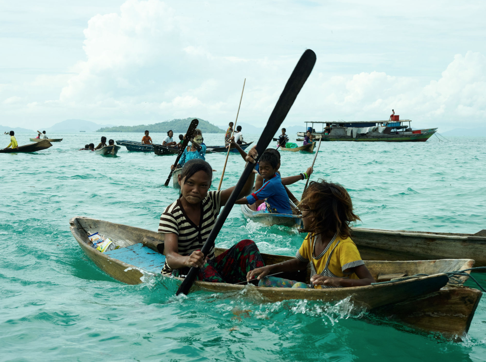
The Bajau people, or “Sea Nomads”, of the Southeastern Asian Islands are renowned as the best divers in the world. The Bajau have been occupying houseboats for generations, living an entirely marine existence as deep-water hunter-gatherers. They spend hours each day underwater, reaching depths of over 200 feet, equipped with no more than weights and wooden goggles. Numerous studies from Tibet looking at physiology and genetics of high altitude societies have raised the question, how are the best divers in the world adapted to their niche?

A recent study compared the Bajau to the neighboring land-dwelling Salaun people, by analyzing DNA, and spleen size. The human body is well adapted to diving; upon breath holding and the sensation of cold water on the face, the body will change its behavior to limit oxygen consumption and contract the spleen to release oxygenated red blood cells into the circulatory system. The Bajau people’s seemingly superhuman diving skills may be explained by physiological differences, like larger spleens.

Researchers took measurements of non–related Bajau (to avoid familial similarities) using ultrasounds and saliva samples to analyze spleen size and DNA, respectively. As a means of comparison, the scientists took similar measurements from the Salaun people of the same area, who have been identified through DNA to be amongst the most genetically similar to the Bajau, but live a grounded existence with little interaction to the marine environment.

The results show that the Bajau people did have significantly larger spleens than the Salaun people and DNA analysis confidently informed a specific gene related to spleen size and thyroid hormones that was expressed at significantly greater levels in Bajau than Salaun people. This is the physiological and genetic difference the researchers were looking for.

So does that mean professional divers have larger spleens than people who can’t swim? Not necessarily. These differences are the result of natural selection. For thousands of years the Bajau have occupied houseboats, traversing the seas of Southeast Asia, living off of materials and food gathered from deep sea floors, using only simple tools. Over so many years, generations, and deep-water dives, these people have evolved. The environment changed their bodies and their genetics to make them better divers. Imagine how your ancestor’s lives adapted you.


Source:

Ilardo MA, Moltke I, Korneliussen TS, Cheng J, Stern AJ, et al., (2018) Physiological and Genetic Adaptations to Diving in Sea Nomads. DOI: 10.1016/j.cell.2018.03.054

Is It Cancer? Check Your Ink

Mouse tattoos
Calcium detecting tattoos in mice

In recent years increased research has been done on the medical applications of tattoos, they have proven effective in delivering drugs, administering vaccines, and working as blood sugar level sensors in conjunction with skin based electronics. New research out of Switzerland, published in Science, has essentially invented a tattoo that can detect cancer. The tattoo “ink” is injected under the skin, and only becomes visible when it detects elevated calcium levels in the blood, an indicator of breast cancer, colon cancer, lung cancer, and more.


The procedure was developed and demonstrated in mice using reprogrammed calcium detecting cells to produce melanin, a black pigment which color moles and freckles. Thus the tattoo would be invisible when administered, but would appear as a large black dot if blood calcium levels became consistently elevated, as in many types of cancer. Perhaps the greatest difficulty in cancer treatment is diagnosis; by the time a patient notices symptoms and seeks medical attention, the disease may have already begun to grow aggressively. By administering these dormant monitors in people, particularly those at a high risk, cancer can be detected as soon as physiological changes begin.


The tattoo ink is actually made from calcium detecting cells that humans use to regulate calcium in the blood. Calcium is an essential compound for maintaining functions like cell growth, cell death, cell movement and more. But its concentration is precisely regulated, if there is too little the bone marrow will produce more, if there is too much, the bones will stop producing it. So when calcium does become elevated it is usually corrected in a few minutes. But certain cancers can inhibit the hormone that acts to regulate calcium production, and so calcium levels remain consistently elevated.


Though there are other pathologies that can cause elevated calcium levels, the tattoo worked well in mice to detect certain cancers before they became symptomatic. It has also been shown to be resilient to short term fluctuations in blood calcium levels and would be especially useful for patients who display risk factors for these cancers such as those with a diagnosed parent, a history of smoking, or work in hazardous environments.


But even if it is best used in niche applications, this represents a dramatic shift towards an entirely new field of diagnostic tests. Human beings can already have their DNA analyzed and identified for specific disease risk factors, if similar tattoos can be created for Celiac Disease, Parkinsons, Alzheimers, and the rest, then medical attention can be sought and treatment strategies implemented when the disease has only just taken hold, when it is at its most treatable.


Tastanova A, Folcher M, Müller M, Camenisch G, Ponti A, et al., (2018) Synthetic biology-based cellular biomedical tattoo for detection of hypercalcemia associated with cancer. DOI: 10.1126/scitranslmed.aap8562

They Survived the Holocaust to Battle Cancer

Holocaust memorial Boston

The Holocaust was the genocide of millions of Jews by the Nazis between the years 1939 and 1945. Researchers at Ben-Gurion University of the Negev in Israel published a study in 2018 that found a higher rate of cancer occurrence in Holocaust survivors in Israel compared to those who did not experience the Holocaust. There is very little information regarding cancer in this group of individuals. All legal decisions concerning financial assistance for Holocaust survivors fighting cancer is based only on one former study. Keeping this in mind, the researchers conducted a comprehensive study that took into account misleading cancer risk factors like age, obesity, diabetes, and smoking.

The study was conducted on 294,543 individuals who were Holocaust survivors based on recognition status as defined by the Holocaust Survivor Benefits Law. Data analysis was done on some of the most prevalent cancers in Israel, cancers of the lungs, colon, bladder, melanoma, breast, and prostate. Analysis for the last two cancers were conducted only on women and men respectively. The occurrence of all six types of cancer was high among Holocaust survivors. The RR, ratio of risk in the exposed (Holocaust survivors), compared to the unexposed (no Holocaust experience) was high for all cancer types. As an example, lung cancer, bladder cancer, and melanoma all showed an RR of 1.2. Based on the fact that the the study took other additional risk factors into consideration, researchers concluded that being a Holocaust survivor is an independent risk factor for all six types of cancer that were explored.

Holocaust survivors who were enrolled in the study were born in European countries under Nazi occupation before 1945, immigrated to Israel after 1945 and were alive in the year 2000. Researchers who conducted the study explain the results as possible consequences of  extreme survival conditions endured during the Holocaust.

Starvation endured by the survivors over extended periods of time could be one explanation for these results.

In addition, continued exposure to toxic wastes due to the proximity of incinerators in concentration camps and, lengthy exposure times to sunlight and resulting UV rays could also be possible reasons.

There are, however, a few shortcomings to this study to be noted. Study participants, despite having immigrated to Israel from a certain country, could have been born in a different one, thus affecting their experience regarding the Holocaust. Furthermore, data analysis was done only based on six common cancer types, when there are many others to be considered. The risk factors based on lifestyle, such as alcohol consumption and exercise habits during the study were not examined. In addition, the Holocaust survivors who died before the year 2000 were not included in the study. This prevents us from taking into consideration their cause of death, which could have shown a higher rate of factors other than cancer.

Taking these advantages and shortcomings into consideration, it can be concluded that the study provided significant results that can be influential in changing the treatment and benefits provided to Holocaust survivors battling cancer in Israel.

Reference:

Ben, D.R., Biderman, A., Sherf, M., Zamstein, O., Dreiher, J. 2018 Elevated cancer risk in Holocaust survivors residing in Israel: A retrospective cohort study. European Journal of Cancer 95: 85-92

Link to article

Link to Image

Johnny Bravo’s square jaw vs Popeye’s round jaw: A scientific explanation

From left to right, popular cartoon characters Ferb, Phineas, Popeye, and Johnny Bravo.

Do you ever scratch your head and roll your eyes at the ridiculous shapes of cartoon characters? While it can’t be confirmed that Phineas and Ferb have legitimate jaw bones, there may be a scientific explanation to Popeye’s rounded jaw to Johnny Bravo’s square jaw.

A recent study published online in Scientific Reports on April 16, 2018, validated a significant association between mandibular shape and jaw muscle cross-sectional size. In other words, researchers found that thicker jaw muscles produced broader, bigger, and more rectangular jaw bones.

Previous studies have shown that craniofacial skeletal form, or the structure of the bones of the face and jaw, is influenced by mechanical loading. Just as your leg bones get stronger from running, and arm bones get stronger from lifting weights, jaw bones get stronger from chewing. The specific shape of the mandibular bone is also determined by the forces applied to it throughout development. So, you might get your dad’s square jaw through genetics, but you also have a square jaw because of the foods you eat regularly.

Jaw muscles
The temporalis muscle and master muscle of the skull. Credit: Sella-Tunis et al. Labels added.

Scientists at Carmel Research Center in Israel measured jaw shapes and jaw muscles in 382 adult patients by utilizing CT scans. These scans allow for visualization of both bone and muscle, and they specifically looked at (1) the temporalis muscle, which is a large, round muscle that reaches from the side of the skull to the side of the face, and (2) the masseter muscle, which stretches from the lower jaw to the upper jaw.

Jaw bone
Comparison of jaw bone size and shape. Credit: Sella-Tunis et al. Labels added.

Independent of gender and accounting for relative size of individuals, researchers found that larger jaw muscles resulted in a wider ramus, a bigger coronoid projection, a more rectangular base, and a more rounded basal arch. Alternatively, smaller muscles produced a skinnier ramus, a smaller coronoid projection, a narrower and angled base, and a more triangular basal arch (see picture above).

This research can be used in anthropology contexts. Researchers suspect that  hunter-gatherer populations had harder diets, comprised of nuts and meat, which generate larger muscles and produced a stronger jaw line, while agricultural groups that ate more vegetation had skulls that resembled the jaws with smaller muscles. According to this data, it is plausible that Popeye’s spinach diet led to the growth of his softer, rounder jaw, and I would guess that Johnny Bravo is a fan of tougher foods.

Source:

Sella-Tunis, T., Pokhojaev, A., Sarig, R., O’Higgins, P., & May, H. 2018. Human mandibular shape is associated with masticatory muscle force. Scientific Reports 8. [doi: 10.1038/s41598-018-24293-3].


ADHD Among College Students, Let’s Be Aware

ADHD

Attention-deficit/hyperactivity disorder (ADHD) is a neurodevelopmental and psychiatric disorder found in all age groups. Past studies have observed simultaneous occurrence of ADHD and other psychiatric disorders in children and adults. To bridge the gap between the two age groups, researchers at the University of North Carolina in Greensboro published a study in 2018 that examining the patterns of non-ADHD psychiatric disorders among 443 emerging adults (1st-year college students aged 18-22 yrs) with and without ADHD. Clinical assessment using multiple methods like interviews, observations, tests and expert reviews showed a significantly high rate of such disorders among students with ADHD.

Comorbidity is the term used to describe the simultaneous occurrence of two chronic diseases or disorders among individuals. The effects of comorbidity of psychiatric disorders and ADHD on the educational and social functioning of college students is still unclear to the scientific community due to the limited number of studies and their varying results.This 5-year investigation was conducted to assess comorbidity in 1st-year college students with ADHD compared to a non-ADHD group. The rate of one current non-ADHD diagnosis was significantly higher for the ADHD group (55%) compared to the non-ADHD group (11%). Amongst the diagnoses were depressive disorders, anxiety disorders, trauma- and stress-related disorders, and learning disorders. Interestingly enough, this rate was higher than those observed among children, but lower than in adults. This raises the possibility of continuity in the development of comorbidity with age among individuals with ADHD.

The study also analyzed the potential influence of ADHD type, gender, and ethnic/racial diversity status on comorbidity in ADHD patients. ADHD can be divided into predominantly hyperactive-impulsive, predominantly inattentive, or combined. Due to the lack of significant sample numbers for the first group, only the latter two groups were compared. Among the two, the combined group showed a much higher rate of having one current comorbid condition. Based on gender, women with ADHD were more likely to have a comorbid disorder compared to men. However, there was no difference found amongst the various ethnic and racial groups. This can be accounted to the disparity in the number of individuals with different diversity status.


women with ADHD
Within the ADHD group, women showed a higher rate (68.4%) of having a comorbid disorder compared to men (40.6%)

Further research is necessary since the study did not specify the possible reasons for the comorbidities associated with ADHD, for example, whether it was increased risk or simply overlapping symptoms. In addition, the samples were all from college campuses in the US, so it cannot be generalized to students in other parts of the world. The effects of comorbidity on educational and social functioning were also not established. However, based on the results, we can agree that there is a distinctly high rate of comorbidity among students with well-defined ADHD in the US. This greatly increases the risk of college students with ADHD experiencing psychosocial difficulties.

In light of these facts, there is a huge necessity for parents and high school educators to be aware of such comorbidities and work together with the students to receive proper care during the transition into college. Furthermore, colleges and universities that provide clinical and academic support services must be aware of the possibility of comorbidities among students with ADHD and  provide proper evaluations and treatment services. This study has helped bridge the gap between comorbidities in ADHD patients of various age groups. For the development of clinical assessment and treatment, future studies need to find proper reasoning for the association between ADHD and co-occurring psychiatric disorders.

Reference:

Anastopoulos, A.D., DePaul, G.J., Weyandt, L.L., Morrissey-Kane, E., Sommer, J.L., Rhoads, L.H., Murphy, K.R., Gormley, M.J., Gudmundsdottir, B.G. 2018 Rates and Patterns of Comorbidity Among First-Year College Students With ADHD. Journal of Clinical Child & Adolescent Psychology 47(2):236-247

Link to article

Link to image 1, Image 2

New skin sensors collect health data, study reports

Body temperature map
Body temperature map. All temperatures were collected by skin sensors, and transmitted to a computer to generate an image. Credit: Han et al.

The era of health tracking is upon us. The ability to track details about our moment-to-moment health has grown exponentially in recent years, from Fitbits logging our sleep patterns and heart rates, to ‘Smart Pills,’ which transmit a signal from a microchip imbedded in a swallowable tablet to an iPhone in order to track compliance with a pill regimen. Recently, a research group working at Carle Hospital at the University of Illinois took the concept of health data to the next level by tracking body temperature and pressure in bedridden patients and sleep study patients. Instead of tracking data from one point, like the wrist, they collected data from the entire body, which is a progressive approach in health tracking technology.

The newly developed skin sensors are thin, soft, wireless, and made of silicon. They have been shown to adhere to the skin without causing irritation, and they are waterproof, so they can collect data even in the shower. In testing, the skin sensors were placed at various points over the whole body, creating a temperature-map and pressure-map of the wearer.

Skin sensors
Sensors placed on the back, and the back of the arms and legs to collect temperature and pressure data. Credit: Han et al.
Pressure map
Pressure map. Credit: Han et al.

Because they are wireless and battery-free, they can stay on a patient for long periods of time, and can easily travel with a patient between wards in a hospital, or from an intensive care facility to a nursing home. They can stay on during medical treatments and during physical therapy. They collect and transmit data to a computer every 3 seconds, so a continuous digital picture of temperature and pressure can be recreated online.

I personally don’t the idea of swallowing a microchip along with my medication, but this new study utilizing skin sensors has incredible implications for medicine. The study, published online in Science Translational Medicine in April 2018, explained that since body temperature naturally fluctuates between day and night, the sensors can be used to measure circadian rhythms. In this case, researchers tested the sensors in a sleep clinic.

The researchers also used the sensors to measure prolonged pressure on the body in bedridden patients. Pressure ulcers, also known as bed sores, are a concern for patients in long-term recovery who spend the majority of their time lying down. Bony areas of the body, like shoulder blades and the buttocks, can develop these bed sores, or irritations, from staying in one position too long. The skin sensors, strategically placed in high-risk areas for bed sores, can detect when the pressure reaches a harmful level.

Source:

Han, S., Kim, J., Won, S.M., Ma, Y., Kang, D., Xie, Z., … Rogers, J.A. (2018). Battery-free, wireless sensors for full-body pressure and temperature mapping. Science Translational Medicine, 20(435). [Published online 04 Apr 2018]. DOI: 10.1126/scitranslmed.aan4950.

Dietary Fiber Decreases Diabetic Difficulties

Every human digestive system is home to trillions of bacterial cells, known as a microbiota. In fact, there are more bacteria in a person’s digestive tract than there are human cells in their entire body. These bacteria play an essential role in digestion, breaking down otherwise indigestible nutrients, into compounds that humans can use. This study finds that increased consumption of fiber can lead to a dramatic reduction in symptoms for patients with Type 2 Diabetes.


Although dietary fiber is indigestible to humans, it is fermentable by the gut microbiota. The gut bacteria ferment the dietary fiber and break it down into short fatty acids, which are essential for moderating glucose, or sugar, concentration, and maintaining proper human health. It is when these fatty acid molecules are lacking that the effects of Type 2 Diabetes are most severe.


Type 2 Diabetes is characterized by an imbalance in glucose concentration in the blood. By increasing short chain fatty acids, excess glucose can be metabolized (broken down and utilized) and the effects of Type 2 Diabetes are greatly diminished. It should come as no surprise that a dietary solution can be offered for a disorder that is largely the result of dietary imbalances, but this study restates the critical importance of the gut microbiota in digestion, and diabetes research. Because though many essential nutrients are indigestible to by humans, there is a lot more at play in digestion than the human physiology.


This study was published in Science Magazine, and serves as a reminder that the gut microbiota is a complex ecosystem that differs greatly from person to person depending on a variety of factors. The relationship between gut bacteria and humans is a mutually beneficial one, as the human consumes fiber, which the bacteria breaks down into usable compounds, which both bacteria and human can utilize. This research may serve to shift the focus in diabetes treatment from dietary glucose, to a more comprehensive understanding of digestive processes.


Zhao L, Zhang F, Ding X, Wu G, Lam YY, et al., (2018) Gut bacteria selectively promoted by dietary fibers alleviate type 2 diabetes. Science 359 (6380), 1151-1156

What does ADHD look like in the brain of a preschooler?

According to the National Survey of Children’s Health from 2016, 9.4% of children in the United States have been diagnosed with ADHD, almost 1 in 10 children. ADHD, or attention-deficit/hyperactivity disorder, is a neurological disorder characterized by impulsive behavior, different attention patterns, restlessness, and disorganization. You might hear a coworker casually claim, “I can’t focus on these emails.. my ADHD is acting up!” However, ADHD is not something that comes and goes. It is a different way of thinking, which stems from abnormal brain developmental patterns.

Although ADHD can manifest as early as 4 years old, most studies have only analyzed older school-age children. A new study published online in March 2018 in the Journal of the International Neuropsychological Society recruited a group of 4-5 year olds, including 52 children exhibiting ADHD symptoms and 38 children without ADHD symptoms to use as a comparison group. With consent from both the parents and children of course, they performed MRI scans to get a look at their brain structure.

Brain scan
A brain scan showing gray matter borders (cell bodies) with white matter in the inner regions of the brain (cell axons); Credit: Wikipedia Commons.

Overall, the researchers found that some regions of the brain had a smaller volume in children with ADHD, compared to the group of children with no ADHD symptoms. In particular, gray matter volumes were decreased. Gray matter, named for its natural brownish-gray color, is tissue comprised of the cell bodies of neurons in the brain and spinal cord. A neuron cell has a central body, and a long axon “branch” which sends messages to other neurons. The neuron cell bodies tend to congregate together in the brain and arrange themselves as “gray matter.” The axons also form groupings and are visualized as white matter.

In the brains of children with ADHD, the researchers noticed that the gray matter volume was reduced most significantly in subregions of the right frontal lobe and the left temporal lobe of the brain, and greater losses in volume corresponded with greater severity of ADHD symptoms. These brain areas with smaller gray matter volume are involved in inhibitory control (for example, preventing one’s self from blurting out an answer instead of raising a hand in class), working memory (for example, remembering the question on a worksheet while in the middle of writing an answer), planning (for example, deciding to clean up the desk, then turn in homework, then put books in backpack in that order), and response control (for example, correctly following a teacher’s directions).

Brain schematic
A schematic of the brain showcasing the frontal lobe and temporal lobe, each of which play a role in ADHD symptoms; Credit: w:User:Washington irving, Wikipedia Commons.

Previously, gray matter volume differences have been assessed in older children, but this study demonstrates that brain structure developments are discernible in children as young as four. The gray matter volume of another brain area, the anterior cingulate cortex, which plays a role in attention, decision-making, and impulsivity, has been evaluated in other studies. In older children with ADHD, there is a reduction in volume of the anterior cingulate cortex, but there was no difference between groups in the 4-5 year olds, suggesting that neural development is transpiring during the course of several years.

Scientists are gaining a better understanding of developmental trajectories of  ADHD with this kind of research. The hope is that these research studies will one day shed light on what triggers the differences in gray matter volume. These neurological differences are believed to be shared by Albert Einstein, Walt Disney, and John F. Kennedy, who also had ADHD symptoms. With this knowledge, we can gain a greater appreciation of what makes us who we are.

Sources:

Children and Adults with Attention-Deficit/Hyperactivity Disorder (CHADD). (2018). General Prevalence. CHADD: The National Resource on ADHD. Retrieved Apr 3, 2018 from http://www.chadd.org/Understanding-ADHD/About-ADHD/Data-and-Statistics/General-Prevalence.aspx.

Growl, J.M. (2018). Famous people with ADHD. PsychCentral. Retrieved Apr 3, 2018 from https://psychcentral.com/lib/famous-people-with-adhd/.

Jacobson, L.A., Crocetti, D., Dirlikov, B., Slifer, K., Denckla, M.B., Mostofsky, S.H., & Mahone, E.M. (2018). Anomalous brain development is evident in preschoolers with attention-deficit/hyperactivity disorder. Journal of the International Neuropsychological Society, First View [Published online]. https://doi.org/10.1017/S1355617718000103.

Mayo Clinic Staff. (2018). Adult attention-deficit/hyperactivity disorder (ADHD). Mayo Clinic. Mayo Foundation for Medical Education and Research. Retrieved Apr 3, 2018 from https://www.mayoclinic.org/diseases-conditions/adult-adhd/symptoms-causes/syc-20350878.


Health Screening for Military Veterans: Do It Right Now!

Marine Corps

Alcohol use disorder (AUD) and post traumatic stress disorder (PTSD) are two of the most common disorders among U.S. military veterans that often occur concurrently. Although both disorders are individually associated with poor health and wellbeing, past studies have shown that the rates of physical and mental health problems are higher in patients with co-occurring AUD and PTSD. Researchers from the National Center for PTSD and the VA San Diego Healthcare System published a study in 2018 that examined the burden associated with the co-occurrence of AUD/PTSD relative to either disorder occurring alone among US military veterans.

The study was conducted using data from the the National Health and Resilience in Veterans Study, which surveyed a nationally representative sample of 3,157 US veterans aged 21 and older. Overall, the sample group consisted of 14.8% probable current AUD, and 16.4% current PTSD, but only 2.8% comorbid AUD/PTSD. Comorbid is a term used when two or more disorders or diseases occur at the same time. As part of the study, veterans were asked about their primary source of healthcare and their current mental health care situation.

The study assessed AUD and PTSD along with probable depressive and anxiety disorder, physical and mental functioning, quality of life, and cognitive functioning.

Results showed that, patients with comorbid AUD/PTSD or PTSD only were more likely to have co-occurring depression and anxiety, and current suicidal ideation compared to veterans with AUD only. Additionally, they also scored lower on mental, physical and cognitive functioning, and quality of life measures. Compared to PTSD only, the comorbid group was, again, much more likely to have attempted suicide in their lifetime. The difference in the rates of depression and anxiety between the two groups was also very high. However, some similarities between the two groups suggests that the increased burden associated with PTSD/AUD is largely driven by problems associated with PTSD. This may have been because the AUD/PTSD group was very small and there was unevenness in the size of the diagnostic groups.

There were also some other limitations associated with the study, which could have brought about discrepancies in the final results. The prevalence of AUD could have been underestimated because AUD is primarily associated with young adults, but the study mostly consisted of older white men. Moreover, suicide attempts were asked in terms of entire lifetime, so it is not certain if they had occurred before or after military service. Nevertheless, the overall findings reflect the mental and physical health burden associated with co-occurring AUD and PTSD.

The results stress the importance of screening veterans for AUD and PTSD given that less than half of veterans with these disorders are receiving mental health treatment. It also highlights the need for further research to understand how to reach out to and engage these veterans in screening, assessment, and treatment for such disorders.

Reference: Norman, S.B., Haller, M., Hamblen, J.L., Southwick,S.M., Pietrzak, R.H. 2018 The Burden of Co-Occurring Alcohol Use Disorder and PTSD in U.S. Military Veterans: Comorbidities, Functioning, and Suicidality. Psychology of Addictive Behaviors 32(2): 224-229

Link to article

Link to image

4 Herbal Supplements for Anxiety and Depression

Passionflower image
Passionflower; Credit: Martin Thomas, Creative Commons
Saffron flowers
Saffron flowers; Credit: Ioulrc, Creative Commons
Chaste berry
Flowering chaste-tree; Credit: Tatters, Creative Commons
Lavender; Credit: Amanda Slater, Creative Commons


According to a recent study, one-third of cancer patients suffer from anxiety, depression, or adjustment disorder in the months following their diagnosis. As a result, many of them add prescription anxiolytic (anti-anxiety) and antidepressant drugs to their cocktail of chemotherapy, radiation therapy, anti-coagulants, and antibiotic drugs.

The problem is that some of these anxiolytic and antidepressant drugs interact with cancer treatments and are less effective in cancer patients. They also trigger a horde of negative side effects that compound the side effects of regular cancer treatments, including seizures, headaches, and addiction.

A group of scientists at the Memorial Sloan Kettering Cancer Center in New York City decided to take a closer look at alternative herbal remedies to treat anxiety and depression in cancer patients. Research done on herbal supplements and plant extracts has been scarce, so the scientists examined a collection of studies completed between 1996 and 2016. By gathering and organizing the data, they noticed that not only are several alternative remedies helpful in ameliorating anxiety and depression, they also counteract aversive effects of chemotherapy and even combat cancer themselves. While not a perfect substitute, the following herbs have promising potential:

1. Extracts of saffron, a spice derived from a Middle Eastern flower, may be able to treat mild to moderate anxiety about as well as fluoxetine (Prozac) and imipramine (Tofranil). It has also been successful in easing anxiety and depression caused by PMS in women.

2. Lavender pills, made from oil of the lavender plant, are able to treat anxiety comparable to the drugs paroxetine (Paxil) and lorazepam (Ativan), but with fewer side effects. Lavender lotions and diffuser oils are often advertised for their calming and relaxation properties, and this holds true for lavender tea and extract drops, which may increase the efficacy of antidepressants citalopram (Celexa) and imipramine (Tofranil).

3. Passionflower, although no better than prescription drugs, seems to perform similarly but with fewer side effects, when compared to oxazepam (Serax) and sertraline (Zoloft). This substance also comes from a flower, which Native Americans have historically used to prevent insomnia.

4. Chasteberry, typically used for PMS symptoms, was compared to fluoxetine (Prozac), and while it didn’t seem to address psychological symptoms of depression, including persistent sadness, hopelessness, and loss of interest, it did alleviate physical symptoms, such as sleep trouble, digestive problems, muscle aches, and headaches.

Overall, researchers found that the herbs are not as potent, but are safer than the prescription counterparts. Clinical trials are needed to further analyze the potential of these herbal supplements and determine their benefits, especially within a oncology context. Because these supplements can be purchased over the counter, physicians don’t always know which supplements their patients are taking. It’s important to discuss an alternative treatment plan with a doctor before use.

Source:

Simon Yeung, K., Hernandez, M., Mao, J.J, Haviland, I., & Gubili, J. (2018). Herbal medicine for depression and anxiety: A systematic review with assessment of potential psycho-oncologic relevance. Phythother Res. [Epub ahead of print]. doi: 10.1002/ptr.6033

Prediabetes Causing Post Stroke Cognitive Impairment

diabetes

Scientists at Wenzhou Medical University in China have established an association between prediabetes and the development of post-stroke cognitive impairment (PSCI). Prediabetes is a state between normal blood-sugar conditions and diabetes mellitus (DM). Cognitive impairment after stroke is fairly common, but varies based on country, race and diagnostic criteria. Prediabetes and DM are both common risk factors of stroke. DM has already been related to PSCI by previous studies, however, as far as the authors’ understanding goes, this was the first study to explore a relationship between PSCI and prediabetes.

Prediabetes is a strong predictor of DM and can be diagnosed by characteristic changes in blood sugar level. Based on these levels, the patients were categorized into diabetes mellitus (DM), prediabetes and non-DM groups. The study evaluated the cognitive function of 201 patients 1 month after a stroke.The study diagnosed 74 of the total 201 patients with PSCI. Data showed the percentage of PSCI patients to be 18.1%, 35.7% and 49.3% respectively for the non-DM, prediabetes, and DM patients.

Analysis based on sex and smoking history surprisingly showed that patients who were female and non-smokers are at a greater risk of PSCI than any other group.

A statistical test conducted to adjust for confounders showed an association between PSCI and prediabetes. Confounders lead to bias that distorts the magnitude of the relationship between two factors of interest. For example, results showed that age and educational level also influenced the effects of prediabetes on PSCI. However, this was found to be untrue after adjusting for confounders.

The study included individuals who had suffered a stroke less than 7 days before admission and were diagnosed using CT scan and MRI. However, it excluded all individuals who had a history of severe depression and other psychiatric disorders or severe nervous system diseases. PSCI was evaluated by experienced neurological physicians who were not aware about the patients’ clinical examination and lab results.

The results were strong, but there were limitations to the study. The use of antidiabetic medicine post stroke was not recorded. Patients with speech and language disorders were excluded, which could have cause biases in the study. Moreover, the time span between stroke and PSCI assessment would have provided more appropriate results if it was longer than one month, for example, 3 to 6 months. Nevertheless, the study proved that prediabetes is a determinant of PSCI and suggests prediabetes patients to maintain normal blood sugar level as a preventive measure.

Reference:

Wang, Q., Zhao, K., Cai, Y., Tu, X., Liu, Y., He, J. 2017. Prediabetes is associated with post-stroke cognitive impairment in ischaemic stroke patients. Elsevier/North-Holland Biomedical Press 1687:137-143

 Link to article

Image 1,  Image 2