What does ADHD look like in the brain of a preschooler?

According to the National Survey of Children’s Health from 2016, 9.4% of children in the United States have been diagnosed with ADHD, almost 1 in 10 children. ADHD, or attention-deficit/hyperactivity disorder, is a neurological disorder characterized by impulsive behavior, different attention patterns, restlessness, and disorganization. You might hear a coworker casually claim, “I can’t focus on these emails.. my ADHD is acting up!” However, ADHD is not something that comes and goes. It is a different way of thinking, which stems from abnormal brain developmental patterns.

Although ADHD can manifest as early as 4 years old, most studies have only analyzed older school-age children. A new study published online in March 2018 in the Journal of the International Neuropsychological Society recruited a group of 4-5 year olds, including 52 children exhibiting ADHD symptoms and 38 children without ADHD symptoms to use as a comparison group. With consent from both the parents and children of course, they performed MRI scans to get a look at their brain structure.

Brain scan
A brain scan showing gray matter borders (cell bodies) with white matter in the inner regions of the brain (cell axons); Credit: Wikipedia Commons.

Overall, the researchers found that some regions of the brain had a smaller volume in children with ADHD, compared to the group of children with no ADHD symptoms. In particular, gray matter volumes were decreased. Gray matter, named for its natural brownish-gray color, is tissue comprised of the cell bodies of neurons in the brain and spinal cord. A neuron cell has a central body, and a long axon “branch” which sends messages to other neurons. The neuron cell bodies tend to congregate together in the brain and arrange themselves as “gray matter.” The axons also form groupings and are visualized as white matter.

In the brains of children with ADHD, the researchers noticed that the gray matter volume was reduced most significantly in subregions of the right frontal lobe and the left temporal lobe of the brain, and greater losses in volume corresponded with greater severity of ADHD symptoms. These brain areas with smaller gray matter volume are involved in inhibitory control (for example, preventing one’s self from blurting out an answer instead of raising a hand in class), working memory (for example, remembering the question on a worksheet while in the middle of writing an answer), planning (for example, deciding to clean up the desk, then turn in homework, then put books in backpack in that order), and response control (for example, correctly following a teacher’s directions).

Brain schematic
A schematic of the brain showcasing the frontal lobe and temporal lobe, each of which play a role in ADHD symptoms; Credit: w:User:Washington irving, Wikipedia Commons.

Previously, gray matter volume differences have been assessed in older children, but this study demonstrates that brain structure developments are discernible in children as young as four. The gray matter volume of another brain area, the anterior cingulate cortex, which plays a role in attention, decision-making, and impulsivity, has been evaluated in other studies. In older children with ADHD, there is a reduction in volume of the anterior cingulate cortex, but there was no difference between groups in the 4-5 year olds, suggesting that neural development is transpiring during the course of several years.

Scientists are gaining a better understanding of developmental trajectories of  ADHD with this kind of research. The hope is that these research studies will one day shed light on what triggers the differences in gray matter volume. These neurological differences are believed to be shared by Albert Einstein, Walt Disney, and John F. Kennedy, who also had ADHD symptoms. With this knowledge, we can gain a greater appreciation of what makes us who we are.

Sources:

Children and Adults with Attention-Deficit/Hyperactivity Disorder (CHADD). (2018). General Prevalence. CHADD: The National Resource on ADHD. Retrieved Apr 3, 2018 from http://www.chadd.org/Understanding-ADHD/About-ADHD/Data-and-Statistics/General-Prevalence.aspx.

Growl, J.M. (2018). Famous people with ADHD. PsychCentral. Retrieved Apr 3, 2018 from https://psychcentral.com/lib/famous-people-with-adhd/.

Jacobson, L.A., Crocetti, D., Dirlikov, B., Slifer, K., Denckla, M.B., Mostofsky, S.H., & Mahone, E.M. (2018). Anomalous brain development is evident in preschoolers with attention-deficit/hyperactivity disorder. Journal of the International Neuropsychological Society, First View [Published online]. https://doi.org/10.1017/S1355617718000103.

Mayo Clinic Staff. (2018). Adult attention-deficit/hyperactivity disorder (ADHD). Mayo Clinic. Mayo Foundation for Medical Education and Research. Retrieved Apr 3, 2018 from https://www.mayoclinic.org/diseases-conditions/adult-adhd/symptoms-causes/syc-20350878.


The rising cost of poor diet: ADHD symptoms in children

A handful of Halloween candy later, and you are skipping around the room, your hands are fidgety, speech is jittery, and it’s hard to contain your burst of energy. You are probably quite familiar with the sugar rush that affects you this one day a year… two days a year? Three days? Almost every day?

Time and again, nutritional studies and long-term research has shown that poor dietary patterns of children influence neural development and behavior, especially hyperactivity and attention capacities, leading to diagnoses like ADHD.

A study from 2009 followed a group of children over several years and found that for significant increases in “junk food” consumption at ages 4-5, the risk for hyperactivity at age 7 was elevated. Another study, from 2004, demonstrated a prolonged association between artificial food coloring and hyperactivity. While the cause of ADHD is still undetermined, dietary patterns certainly play a role. Not only do high-fat, high-sugar diets cause issues in the developing nervous system, but these diets tend to also be low in valuable vitamins and micronutrients.

A more recent study, published online in March 2018 in European Journal of Clinical Nutrition, showed a positive correlation between a “processed” food pattern, a “snack” food pattern, and ADHD symptoms in children age 3-6. On the other hand, there was a negative correlation between a “vegetarian” food pattern and ADHD symptoms. This study used data collected from over 14,000 preschoolers in Ma’anshan City, China, and was the first large scale study in mainland China to investigate connections between diet and ADHD in children. The prevalence of ADHD symptoms in the group studied was 8.6%.

Researchers asked caregivers and parents to answer questionnaires about the recent food consumption and food choices of their children, and gave the children a Conners Abbreviated Symptom Questionnaire to assess for ADHD symptoms. Then, the researchers allocated five dietary patterns to represent the common answers expressed by the caregivers and parents: (1) “processed,” for fast food, fried foods, preserved fruit, and other high-fat food items, (2) “snack,” for high-sugar foods like sweets, biscuits, cakes, and chocolates, (3) “protein,” for red meat, poultry, eggs, fish, fruit, and rice, (4) “beverage,” for flavored milk, soda, and yogurt, and (5) “vegetarian,” for grains, beans, and fruit or vegetable juice.

Children who had a “processed” or “snack” dietary pattern had a significantly higher likelihood of demonstrating ADHD symptoms, hyperactivity, and attention problems. There was no correlation between ADHD symptoms and the “protein” or “beverage” categories, but the “vegetarian” dietary pattern seems to act as a protector against ADHD symptoms.

This study can not show causation, or that a poor diet causes ADHD. However, it can not be refuted that diet is an influencing factor in development and behavior in children. High-fat, high-sugar foods tend to be cheaper, more accessible, and more conveniently packaged in bags and wrappers for on-the-go, and they don’t need to be refrigerated. They taste good. But, the cost is a rising prevalence of ADHD and similar disorders. The relationship between food and hyperactivity is further evident through studies that have shown that elimination diets and fish oil supplements can reverse the ADHD symptoms. Fatty foods and sugary foods should be eaten in moderation; Halloween should not be occurring every day.

Source:

Yan, S., Cao, H., Gu, C., Ni, L., Tao, H., Shao, T., Xu, Y., & Tao, F. (2018). Dietary patterns are associated with attention-deficit/hyperactivity disorder (ADHD) symptoms among preschoolers in mainland China. European Journal of Clinical Nutrition [Published online March 13, 2018]. doi:10.1038/s41430-018-0131-0.

The Beginning of an End to the Autism-Vaccine Debate?

Autism Awareness

Autism spectrum disorder (ASD) is a developmental disorder of the nervous system. The causes of ASD are yet unknown, but it has been linked to both genetic and environmental factors. Researchers at Keele University in the UK have identified aluminum as a potential cause of autism based on a study conducted in 2017 on brain tissue from people diagnosed with ASD.

Aluminum is used in vaccines to enhance the body’s immune response to antigens (harmful or toxic substances). The vaccine-autism debate is highly controversial, but animal models have linked the use of aluminum in vaccines to ASD. The results of this study on human cells further assert these findings.

Aluminum content was measured in 0.3g tissue samples from different regions of the brain of 5 individuals using atomic absorption spectrometry. This method utilizes the difference in the light absorption capabilities of different atoms to find the chemical composition of samples. The values ranged from 1.20 to 4.77μg/g. Past studies have suggested values ≥2.00μg/g as pathologically concerning and those ≥3.00μg/g as pathologically significant. The results showed at least one tissue in each individual that exceeded the established pathologically significant value.

Some of the values recorded were the highest ever measured (17.10, 18.57 and 22.11μg/g).

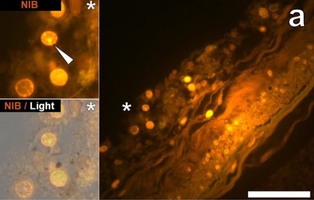
In addition to the concentration, the locations of the aluminum deposits were also examined using fluorescence microscopes. A dye that selectively stains aluminum in cells and human tissues and makes them appear orange or bright yellow was used to view aluminum on the images obtained through the microscope. Deposits were found both inside and outside brain cells. However, the most distinct observation was the presence of metal deposits in the microglia. Microglia are the main immune defense cells inside the central nervous systems and scientists concluded that the deposits seen in them were a direct indication that aluminum had somehow crossed the blood-brain barrier.

fluorescence micrograph
Figure 1 shows the cells in the hippocampus of a 50-year-old male donor used in the study by Mold et al. The white arrow indicated aluminum depositions that were observed via orange fluorescence emission. Hippocampus is the part of the brain considered to be the center of emotion and memory.

Aluminum is toxic to living cells. Although the microglia could remain functional for a certain time period, the metal will eventually show its adverse effects by disrupting this functionality. This directly correlates defective microglia with ASD. In addition to microglia, the study showed aluminum depositions in other tissues from different parts of the brain.

The study also showed great variability in the age groups of donors from 15 to 50 year olds. Initially, the high concentration seen in tissue from a 15 years old donor had greatly puzzled the researchers. However, the evidence of aluminum deposition in the microglia and other intracellular locations ties back to implicate vaccines as a potential cause of ASD and explain how such high amounts of aluminum could have deposited in the brain tissues of a 15 year old boy.

This shows the first ever instance of aluminum concentration measurement in human brain tissues from individuals with ASD. Despite the concrete results, the research was limited due to the lack of a substantial number of subjects and the minimal amount of tissue cells that could be obtained for the study. These factors render the research inadequate by itself to establish ASD as a direct outcome of aluminum deposition from vaccines in brain tissues. However, it is a major stepping stone towards realizing the potential cause of autism spectrum disorder. Now, there is a need for more research to either support or question the results of this study. 

Reference:

Mold, M., Umar, D., King, A., and Exley, C.2018. Aluminium in brain tissue in autism. Journal of Trace Elements in Medicine and Biology 46: 76-82.

Link to article

Link to feature image