Will therapy benefit OCD patients? Computers have the answer.

fMRI machine
Patient being prepared for an fMRI. Credit: Ptrump16, Creative Commons.

Obsessive-compulsive disorder, or OCD, is characterized by unwanted, repetitive thoughts and impulsive, ritualistic actions. For example, a common fear among those with OCD is a fear of germs, which results in repetitive hand-washing. While historically OCD has been difficult to treat effectively, in recent years, modifications to cognitive-behavioral therapy have had more success. Cognitive-behavioral therapy is comprised of a series of sessions between a therapist and patient to identify negative thought patterns and symptoms, and address them through discussion, exposure to stress-inducing stimuli, and practice utilizing alternative coping mechanisms to ameliorate anxiety.

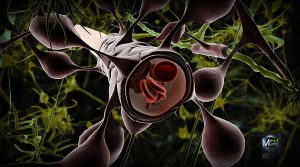
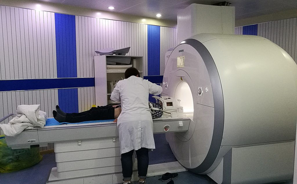
While cognitive-behavioral therapy can be effective, it is time-consuming and does not work for everyone. Using functional magnetic resonance imaging (fMRIs), scientists at UCLA trained a computer analysis system to study the brains of individuals with OCD, and determine which individuals were most likely to benefit from cognitive-behavioral therapy. Their study demonstrated that if an OCD patient were to receive a seven-minute fMRI scan, the computer program could predict the success of cognitive-behavioral therapy for that particular patient, at 67-70% accuracy.

For their study, researchers recruited 42 adults with OCD. All of the participants underwent fMRIs at the beginning of the study. Then, half of the participants attended cognitive-behavioral therapy sessions lasting about 90 minutes per session, five days a week for four weeks. At the end, their brains were analyzed with an fMRI again to detect any differences in structure or brain function. The other half of the participants were put on a four-week waitlist. At the end of four weeks, having received no therapy, their brains were scanned to see if there were any differences simply due to time. These participants received cognitive-behavioral therapy treatment after the four-week waiting period.

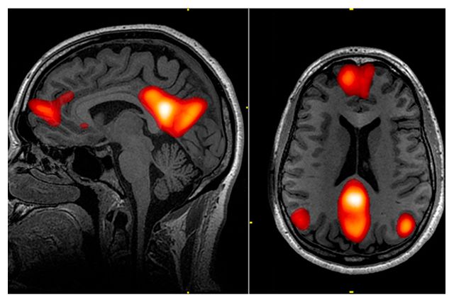
On the fMRI scans, the researchers were especially interested in studying the regions of the brain and their cellular networks that regulate attention, vision, motor skills, memory, self-evaluation, and the abstract sense of “mind-wandering,” or daydreaming, each of which play a role in development of OCD. They utilized mathematical models and computer learning to map differences between the participant’s brains, and match those results with behavior results of cognitive-behavioral therapy. They found that the computer could suggest which patients would benefit from therapy, regardless of individual symptoms or severity of symptoms.

fMRI brain scan
One of the brain networks studied was the default mode network, or DMN, which plays a role in “mind-wandering,” daydreaming, and abstract thought involved in thinking about the self. Regions of the DMN are highlighted in red in this fMRI scan. Credit: Leigh Hopper, UCLA Newsroom.

Widespread use of this predictive method would give therapists more information when deciding the best route of treatment for their patients. In the study, the researchers advocate for this fMRI computer model as a way to allocate time and resources, and direct cognitive-behavior therapy towards patients who are most likely to have success, versus other types of treatment such as medications, inpatient programs, intensive day programs, or group therapy. It is a move towards personalized medicine.

However, more research needs to be done to further advance this technique. Computers alone are not yet adequate to diagnose psychological disorders or comprehend subjective human experience. Furthermore, fMRIs are extremely expensive, and the money going towards fMRI scans could instead be put towards treatment. There is also a risk that those who the computer does not deem fit for cognitive-behavioral therapy miss out on a treatment opportunity that could actually help. While studies like this one advance scientific understanding of disorders like OCD, clinicians should proceed with caution when incorporating new computer-based evaluations that could be wrong and depersonalize the treatment experience.

Sources:

Reggente, N., Moody, T.D., Morfini, F., Sheen, C., Rissman, J., O’Neill, J., & Feusner, J.D. (2018) Multivariate resting-state functional connectivity predicts response to cognitive behavioral therapy in obsessive-compulsive disorder. PNAS [published online ahead of print]. https://doi.org/10.1073/pnas.1716686115.

Hopper, Leigh. 2018. Brain scan and artificial intelligence could help predict whether OCD will improve with treatment. UCLA Newsroom. Retrieved Feb. 5 from http://newsroom.ucla.edu/releases/brain-scan-AI-help-predict-ocd-improve-treatment.

Insight into Pericytes

Blood Brain Barrier and Astrocytes type 1
Blood Brain Barrier. Credit: Ben Brahim Mohammed, Wikimedia Commons

Imagine the vascular system in the brain as a strainer used in cooking. After cooking pasta in a pot of water, you pour the pasta over the strainer, so that it catches the noodles, and the water filters out into the sink. Typically, you want a strainer with small holes, so vegetable pieces or meat pieces cooked with your pasta don’t slip out with the water into the sink.

Similarly, specialized cells called pericytes act as the strainer of blood flow in the brain. These cells contribute to forming the blood-brain barrier, which permits nutrients and oxygen to filter through to feed brain cells but prevents toxins from entering the brain. The pericytes play an active role in managing this exchange. Pericytes also regulate blood flow in the small capillary blood vessels. In other words, they determine the width of the blood vessels and decide how much blood can flow freely.

A recent study published in Nature Medicine on February 5th linked pericyte damage with Alzheimer’s Disease and other forms of dementia. Previously, Azheimer’s Disease and other neurodegenerative diseases were associated with accumulations of TAU proteins, toxic proteins that build up over time and inhibit brain function. Researchers at the University of Southern California now think pericytes are to blame as an earlier marker for dementia, causing issues before TAU proteins even show up.

Researchers used a mouse model to simulate pericyte deficiency in humans, and noticed that damaged pericyte cells let some materials leak out of the blood and into the brain that were not supposed to be there, just like a strainer with holes that are too big and macaroni noodles start plopping into the sink. The leaking material was fibrinogen, a protein that creates blood clots at injury sites. During the healing process, fibrinogen is vital, but in the brain, fibrinogen deposits erode away at the insulation barrier of neurons, called myelin, and disrupt electrical communication from one neuron to another. You might equate fibrinogen as the chunks that get through your strainer, and then clog the drain pipe.

Nerve tracts gradually eroding as the result of damaged pericytes.
Myelin (shown in green and red) gradually erodes away as the result of damaged pericytes.  Credit: Montagne et al.

The alarming discovery was that in the absence of healthy pericytes, fibrinogen leaked into the brain, and the cells that produce myelin, called oligodendrocytes, started to die. By the end of the experiment, 50% of the oligodendrocytes were dying or defective. One hypothesis proposed was that besides directly destroying the oligodendrocytes, fibrinogen also blocks oxygen and nutrients from reaching them, further accelerating cell death.

The scientists are hopeful that their research will initiate new treatments for dementia by focusing on the root of the problem: the damaged pericytes producing leaks in the blood-brain barrier. The senior researcher said, “Perhaps focusing on strengthening the blood-brain barrier integrity may be an answer because you can’t eliminate fibrinogen from blood in humans. This protein is necessary in the blood. It just happens to be toxic to the brain.” With future research, the pericytes may become the primary target for dementia treatment and prevention.

Sources:

Montagne, A., Nikolakopoulou, A., Zhao, Z., Sagare, A.P., Si, G., Lazic, D., Barnes, S.R., Daianu, M., Ramanathan, A., Go, A., Lawson, E.J., Wang, Y., Mack, W.J., Thompson, P.M., Schneider, J.A., Varkey, J., Langen, R., Mullins, E., Jacobs, R.E., & Zlokovic, B.V. 2018. Berichte degeneration causes white matter dysfunction in the mouse central nervous system. Nature Medicine [ePub ahead of print].

Vuong, Zang. 2018. Half of all dementias start with damaged ‘gatekeeper cells.’ USC Press Room. Retrieved Feb. 12 from http://pressroom.usc.edu/half-of-all-dementias-start-with-damaged-gatekeeper-cells/.


Alpha waves, attention, anxiety, oh my!

Neurons firing in the brain.
Neurons firing in the brain (artificial color added). Credit: Picower Institute for Learning and Memory, M.I.T.

In a recent study, published in January 2018, scientists pinpointed a unique characteristic of people who experience trait anxiety–differences in alpha brain wave activity. Usually anxiety is correlated with an absence of alpha waves; in anxious brains, beta waves overpower alpha waves, and over time, this accumulates into feelings of constant stress. Researchers in the Departments of Psychology and Psychological Science at Ball State University found that too many alpha waves can create an equally disruptive imbalance.

The brain is composed of billions of neurons, which communicate with each other through electrical signaling. When multiple neurons fire simultaneously, they produce electrical oscillations, or “waves.” The frequency of these waves depends on the current level of consciousness: brain waves tend to be lower frequency during deep sleep, but high frequency during problem-solving, decision-making, and other tasks requiring complex thinking and concentration.

Alpha waves, which were evaluated in this study, are known to occur when the mind is in a state of relaxation. At any given moment, the brain might elicit more than one type of brain wave, but alpha waves are most widespread during meditation, while daydreaming, and even during prolonged aerobic activity, like a “runner’s high.” However, as soon as we are alerted with a task, faster beta waves take over.

This may not be the case with highly anxious individuals. Researchers used an EEG to measure the alpha brain waves of a group of individuals in a high-trait anxiety condition, analogous with having an anxiety disorder, and a group of individuals in a low-trait anxiety condition, meaning they showed very few anxiety symptoms. Researchers first measured the alpha waves during a resting, relaxed state, and then while the participants completed a response-inhibition test called the Eriksen-Flanker Task.

Researchers found that the highly anxious individuals demonstrated more alpha wave activity in the resting state, compared to the less anxious individuals. But during the Eriksen-Flanker Task, the two groups demonstrated similar levels of alpha wave activity. In other words, at baseline, the highly anxious individuals were essentially more relaxed than typical, so their brains had to make a further jump to get to an alert and focused state.

While this may seem counter-intuitive, the implications for this experiment are that the prevailing alpha waves in the brain of a highly anxious individual suppress processing of external stimuli and information. The individual might then have trouble focusing on specific tasks and thoughts. In conjunction with previous studies, anxiety has been linked to a lack of alpha waves as well as extra alpha waves in a resting state, suggesting that abnormal alpha brain wave activity alters attention and processing in various ways. More research is needed to more clearly understand this phenomenon, but researchers hope this method of measuring alpha waves will become a tool to measure degrees of anxiety in the future.

brain waves
Types of brain waves, as they appear on an EEG. Credit: Slaven Cvijetic.

Sources:

Ward, R.T., Smith, S.L., Kraus, B.T., Allen, A.V., Moses, M.A., Simon-Dack, S.L. 2018. Alpha band frequency differences between lot-trait and high-trait anxious individuals. NeuroReport 29:79-83.

Bergland, C. 2015. Alpha brain waves boost creativity and reduce depression. Psychology Today. Retrieved Feb 5, 2018 from https://www.psychologytoday.com/blog/the-athletes-way/201504/alpha-brain-waves-boost-creativity-and-reduce-depression


Researchers Stimulate the Amygdala to Stimulate Memory

Think back to your first kiss, your soccer championship game, or hearing about the death of a loved one. Do you remember what you were wearing? Do you remember who was there with you and specifically where you were? You might even remember the exact words from what people said around you. These crystal-clear memories are called flashbulb memories, and are processed by the amygdala, a region of the brain associated with regulating emotions and emotional memory.

A new study now reveals that directly stimulating the amygdala can result in improved memory without a combined emotional experience. Participants in a study at Emory University Hospital received brief, low-amplitude electrical stimulation to the amygdala and demonstrated improved declarative memory the next day without any subjective emotional feelings or involuntary emotional responses, such as increased heart rate or faster breathing.

The study, published online in December 2017 in the journal PNAS, took place in conjunction with the Emory University School of Medicine. Epilepsy patients with electrodes already implanted in their brains were recruited to participate, and fourteen individuals took part in the study. Participants were shown numerous neutral images (i.e. a picture of a basketball or a key), and then either given a short stimulation of the amygdala or no stimulation. Immediately afterwards and again the following day, participants were shown more neutral images in a recognition-memory test. The patients who received the stimulation exhibited greater memory retention of images after one day, compared to the control group.

While the participants in the study were simultaneously receiving treatment for epilepsy, they showed substantial memory enhancement due to the amygdala stimulation. One patient, who suffered from brain damage and memory impairment and rarely recognized researchers and physicians, displayed the most memory improvement. Other patients who experienced seizures in between the initial stimulation and the test the following day showed improved memory, presenting evidence that the amygdala stores memories in spite of other neurologically debilitating disorders. None of the patients reported being able to feel the stimulation.

brain
Illustration of the amygdala (blue), hippocampus (orange), and perirhinal cortex (pink). Credit: Cory Inman, Emory University.

Researchers speculate that the amygdala plays a role in delegating non-emotional declarative memory to other structures, namely the hippocampus and the perirhinal cortex. Specific stimulation to the hippocampus and perirhinal cortex to improve memory has been erratic in prior studies, and the amygdala might be the missing link. According to co-author Joseph Manns, “the long-term goal of this research program is to understand how modulation of the hippocampus by the amygdala can at times lead to memory enhancement and at times lead to memory dysfunction, such as that observed in post-traumatic stress disorder (PTSD).”

Regarding the targeted amygdala stimulation, co-author Cory Inman explained, “One day, this could be incorporated into a device aimed at helping patients with severe memory impairments, like those with traumatic brain injuries or mild cognitive impairment associated with various neurodegenerative diseases.” Small deep-brain stimulation implants are already being used to treat Parkinson’s disease. This study may be utilized in future research to develop similar clinical treatments for patients with memory disorders, so that their non-emotional memories like what they ate for last night’s dinner or what they read in a good book, can be remembered the next day.

Amygdala and hippocampus highlighted in brain
Limbic system imbedded in the brain. Amygdala is shown in red and the hippocampus is shown in purple. Credit: Paul Wissmann, Santa Monica College.

Sources:

Inman, C.S., Manns, J.R., Bijanki, K.R., Bass, D.I., Hamann, S., Drane, D.L., Fasano, R.E., Kovach, C.K., Gross, R.E., and Willie, J.T. 2018. Direct electrical stimulation of the amygdala enhances declarative memory in humans. PNAS 115: 98-103.

Emory Health Sciences. 2017. Direct amygdala stimulation can enhance human memory for a day: Preliminary study of time-specific electrical stimulation. ScienceDaily. Retrieved January 31, 2018 from www.sciencedaily.com/releases/2017/12/171218151808.htm.