Silver and Gold ‘nanocages’ might buy early detection

By Sara Braniecki

Researchers from Washington University and Purdue University collaborated to experiment with a new medical imaging technique that they hope will lead to early detection and treatment for cancer patients.  The procedure uses a pulsed laser and tiny metallic “nanocages, as the researchers call them, to create images much clearer than those created using previous techniques.

This composite image shows luminous nanocages, which appear like stars against a black background, and a living cell, at upper left. The gold-silver nanocages exhibit a bright "three-photon luminescence" when excited by the ultrafast pulsed laser, with 10-times greater intensity than pure gold or silver nanoparticles. The signal allows live cell imaging with negligible damage from heating.

The nanocages are injected into the bloodstream, and then laser pulses are shone through the patient’s skin to detect them. The nanocages are small, hollow spheres made of a combination of gold and silver. Both the nanocages are only 40 nanometers wide.  To put that into perspective, this is 100 times smaller than a red blood cell.  The laser shines light that is almost infrared and pulses 80 million times per second.

The procedure illuminates tissues and organs, allowing live cell imaging.  The precision of these images is important for accurate detection and thorough treatment of cancer.

The images produced using this technique provide a much better image than older techniques that used nanospheres made solely of gold.  The new images have greater contrast since there is less background glow of surrounding tissues.  One of the researchers from Purdue University, Ji-Xen Cheng, explains, “This lack of background fluorescence makes the images much more clear and is very important for disease detection.  It allows us to clearly identify the nanocages and the tissues.”

Another advantage of using the nanocages made of both silver and gold is that there is no resulting heat damage in the tissue.  Previously, the image needed to be enhanced to get a clear enough image that was usable.  To enhance the image, clouds of electrons moving in unison had to be induced in the tissue- this resulted in the heat damage.  Since this enhancement is no longer vital, the heat damage does not occur.

The researchers hope that the creation of the nanocages will lead to better detection and treatment.  Washington University researcher Younan Xia, whose team engineered the nanocages, explains that the productions of the nanocages will likely allow researchers “to combine imaging and therapy for better diagnosis and monitoring.”  He also foresees that the nanocages might be used to deliver time-released anticancer drugs to diseased tissue.

Further information.

Wait for Prostate Treatment May Be Over

April 18, 2010

By: Shelly Hwang

Prostate cancer is the second-leading cause of cancer death in men, with men having a one in six chance that they will get prostate cancer in their lifetime. While prostate cancer can be treated with surgery, a new treatment similar to radiation is being tested that may be able to more effectively target proteins on the surface of prostate tumors, providing hope even for patients with advanced prostate cancer.

Human prostate cancer cells can be recognized by overexpression of some proteins on their surface. The abundance of certain proteins provides a way to target these cancer cells by using antibodies. The antibodies will be binded to the isotope 212-lead, which is an altered form of the common element lead. When this antibody is injected into a patient’s veins, it will bind to a tumor’s surface and release particles and radiation that will destroy only the tumor cells.

Researchers in Zhongyun Dong’s laboratory at the University of Cincinnati are getting ready to test this new agent over the course of this year. They will measure how toxic and effective the treatment is in slowing down or blocking cancer cell growth. Then, the treatment will be used in clinical trials with patients with advanced prostate cancer.

Original Press Release

UC Academic Health Center

Time for scabby knees

By Nina Jean-Jacques

Have you ever wondered how you get those scabs after you cut yourself? You may already know that it’s due to blod clotting. More studies on blood clotting found the time it takes for blood to clot can be determined by something in the blood – genes.

Researchers at the University of Edinburgh discovered three genes that are responsible for blood clotting. They performed the study at the Centre for Cognitive Ageing and Cognitive Epidemiology. The researchers used blood samples from a group of people in the Edinburgh area who were aged over 70. These participants are also a part of the Lothian Birth Cohort 1921 and 1936, which has been studying the participants since birth.

The study used a test called activated partial thromboplastin time to measure clotting time and examined thousands of genes. The genes that were found to assist in blood clotting are F12, HRG, and KNG1. These genes are found in healthy people. The researchers on this study are trying to encourage the start of studies on genes of those that suffer from blood clotting disorders such as deep vein thrombosis and some types of stroke. The researchers are all so excited to discover such a large genetic component in clotting.

blood clotting disorder - deep vein thrombosis

Researchers solve viral superinfection mystery

By Liz H. ‘10

Interaction between a T-cell (purple) and another cell of the immune system.

The ability of a common virus known as CMV to cause a “superinfection” and infect humans multiple times has puzzled scientists until recently.  Researchers at the Oregon Health and Science University Vaccine and Gene Therapy Institute have reported the mechanism that CMV uses to evade the immune system and re-infect humans, in a study published in the April 2nd issue of Science.  Their findings shed light on how this virus may be used in the development of CMV-based vaccines for other diseases.

Cytomegalovirus (CMV) does something that not many viruses can do:  it can re-infect people who previously have been infected by CMV and have already developed an immune response to the virus.  This is unusual for a virus, because the immune system usually “remembers” previous infections with viruses and other pathogens and can mount a strong immune response upon re-infection with a specific pathogen.

The researchers studied CMV-infected monkeys in order to understand how the virus overcomes detection by the immune system.  They discovered that CMV avoids a special type of white blood cell called CD8+ T cells, which are responsible for killing cells that are infected with a pathogen.  CD8+ T cells recognize infected cells by small molecules on the exterior surface of infected cells known as MHC I.  These molecules display small pieces of an invading pathogen and present them to the T cells.  This presentation of infectious material ultimately signals T cells to start destroying infected cells.

In order to evade these T cells, CMV makes proteins that interferes with the presentation of viral pieces by MHC I molecules and stops them from recruiting T cells to infected cells.  “In essence, CMV is able to cut off an infected cell’s call for elimination. This allows CMV to overcome this critical immune barrier during re-infection,” explains author Klaus Frueh.

The study has interesting implications in the design of CMV-based viral vaccine vectors.  Viral vaccine vectors contain a modified, harmless virus that carries a vaccine for another pathogen to the body.  Although the body develops immunity to the pathogen, it also develops immunity to the viral vector, which means that the viral vector can only be used for one type of vaccine.  Since CMV does not elicit an immune response upon re-infection, it makes an attractive vaccine vector candidate that could potentially carry vaccines against other pathogens, such as HIV, hepatitis C, tuberculosis, and malaria.

CMV is a member of the herpesvirus family and infects 50-80% of the US population by age 40.  Most people do not have any symptoms of CMV infection and do not become ill.  But for those with weakened immune systems, including infants and the immunocompromised, CMV can cause serious complications.  With this new understanding of how CMV evades the immune system, scientists may be able to start utilizing the virus for the benefit of human health.

Want more information?

CMV infection

Press Release

Nano probes: the future of drug delivery?

by Johnathan Nieves ’11 

Scientists have developed an extremely small probing device that is capable of binding to a cells surface and eavesdropping on its internal electrical activity. This may help to provide insight into how cells communicate and how they respond to medication delivered through the probe.    

Ever think we could spy on a cell? We have been able to for almost thirty years now, but a new technique is purported to no do it substantially better. Stanford Researchers recently (March 30) published a paper describing their success in developing a nanometer-scale probe capable of binding and becoming a part of a single cell’s membrane.  The paper, published in Proceedings of the National Academy of Science, offers insight into the ability for researchers to eavesdrop on the inner electrical activity of individual cells. The use of the nano probe as a conduit for inserting medication into a cell’s interior is also being cited by the Stanford researchers.    

The study, spearheaded by Nick Melosh and Benjamin Almquist, focused on designing a probe in a way that allowed it to mimic a component of the cell membrane. The cell membrane, or cell wall, is the outermost encapsulating structure of a cell that protects it from the outside environment. The key to the probe’s easy insertion and the great affinity it has for the cell membrane is due to its engineering. The probe was engineered in a way that allowed it to mimic a type of cell membrane gatekeeper protein – a molecule naturally found in the cell membrane that regulates what enters and exits the cell.      

Image depicting the nano-probe binding to the cell membrane. (Credit: Benjamin Almquist, Stanford University)

“What we have done is make an inorganic version of one of those membrane proteins, which sits  in the membrane without disrupting it,” said Melosh. “The probes fuse into the membranes spontaneously and form good, strong junctions there.” The attachment is so strong, “we cannot pull them out. The membrane will just keep deforming rather than let go of the probes.” The 600-nanometer-long probe has integrated so well into membranes that the researchers have dubbed it the “stealth” probe.    

Current methods involved in cell probing are limited in that they only allow access to the cell for few hours. Additionally, the methods are extremely destructive and damaging to cells. Melosh and Almquist are the first to implant a cell probe with very little damage to the cell.      

Up to now, poking a hole in a cell membrane has largely relied on brute force, Melosh said. “We can basically rip holes in the cells using suction, we can use high voltage to puncture holes in their membranes, both of which are fairly destructive […]; many of the cells don’t survive.” That limits the duration of any observations, particularly electrical measurements of cell function.    

“Ideally, what you’d like to be able to do is have an access port through the cell membrane that you can put things in or take things out, measure electrical currents … basically full control,” commented Melosh. “That’s really what we’ve shown – this is a platform upon which you can start building those kinds of devices.”    

Melosh and Almquist are currently working with human red blood cells, cervical and ovary cancer cells to demonstrate the functionality of the probes in living cells.    

To view the press release pertaining to this article, click here.

Tricky tumors grow and spread

The tumor has transformed its outer layer to mimic lymph node tissue to avoid detection by the immune system.

By Sara Braniecki

Researchers from Switzerland’s Ecole Polytechnique Fédérale de Lausanne (EPFL) published a study in the March 2010 issue of Science explaining how tumor cells avoid being destroyed by the immune system.  The researchers believe that knowing how cancerous cells avoid the body’s immune system could lead to a future understanding of how to use the body’s natural defense mechanism to destroy cancerous cells.

Tumors make themselves and the surrounding area seem perfectly normal by disguising themselves as lymph nodes, which are a key part of the immune system that filters the blood and traps foreign particles.  Due to the disguise, the immune system is not phased and has no reason to take any destructive action on the cancer cells.

The researchers focused on a protein in genuine lymph nodes that attracts cells and instructs them to carry out defensive functions for the body.  Some tumors make their outer layer, with which the immune system would come into contact, appear as lymph node tissue by secreting this protein that the researchers were studying.  Since the tumors secrete the protein, they attract immune cells.   The immune cells are tricked into thinking the tumor is healthy rather than foreign.  Thus the tumor is not destroyed by the immune system, allowing it to grow and spread.

According to one of the researchers, Jacqui Shields, the concept that tumors mimic lymphoid tissue to alter the host’s immune response represents a new understanding of tumors’ interactions with the lymphatic system.  This will possibly open up a new area of study, and hopefully open up new understanding for cancer therapy.

Further information.

Time to Get Paranoid about your Thyroid

By: Shelly Hwang

April 3, 2010

Most young adults from the ages of 18 to 44 don’t give much thought to their thyroid, but a study published earlier this month in Stroke: Journal of the American Heart Association reveals increased risk of stroke in young adults with an overactive thyroid.

So what exactly is an overactive thyroid? Hyperthyroidism, or an overactive thyroid, is a condition that causes overproduction of thyroid hormone, which increases metabolism and causes sweating, diarrhea, weight loss, and nervousness. Hyperthyroidism is common, affecting about 0.5-2% of the worldwide population, particularly young adults. The study shows an association between hyperthyroidism and ischemic stroke, which is the most common type of stroke caused by blocked arteries in or leading to the brain.

The study compared data on 3,176 young adults diagnosed with hyperthyroidism between January 1998 and December 2001 and 25,408 patients without thyroid disease, with the average age being 32 years. After five years, 198 of the 28,584 patients developed ischemic stroke (0.7%), with 1% of the hyperthyroidism patients and 0.6% of the comparison group having a stroke. After accounting for many factors such as age, gender, high blood pressure, diabetes, and an irregular heart rhythm called atrial fibrillation (AF), the risk of hyperthyroidism patients having a stroke was 44 percent higher than those without hyperthyroidism.

In adults over the age of 60, Hyperthyroidism is known to be associated with AF, which occurs when the heart beats irregularly and ineffectively and can lead to a stroke. However, the risk of stroke in younger people with hyperthyroidism has not been previously studied. This study could lead to a new screening process for young adults to help lower risk of developing a stroke sooner than expected.

Press Release

American Stroke Association

History of Smoking Reduces Risk of Parkinson’s Disease

By Kelly Lohr

Believe it or not, a new study has shown that a history of cigarette smoking may actually benefit your health.  Over the last decade, Honglei Chen led a study out of the National Institute of Environmental Health Sciences in Research Triangle Park, North Carolina examining long-term health effects of the habit.  Over 300,000 AARP members between the ages of 50 to 71 were surveyed about lifestyle choices over a ten-year period.  Of these subjects, 0.05% of the individuals developed Parkinson’s disease.

Parkinson’s disease is a neurodegenerative disorder characterized by the breakdown of cells which release the neurotransmitter dopamine in a brain area known as the substantia nigra.   Typical symptoms of Parkinson’s include uncontrollable muscle movements, poor posture, and rigidity.  Of the participants from Chen’s study, it was found that current smokers reduced their risk of Parkinson’s disease by 44% as compared to non-smokers.  Previous smokers who had quit reduced their risk of Parkinson’s by 22%.

Interestingly, the risk of developing Parkinson’s disease did not change based on how many cigarettes a person smoked per day.  Instead, the length of the history of smoking was correlated to reduction in disease risk.  Those who smoked for at least 40 years were 46% less likely to develop Parkinson’s, whereas those who smoked between 30 and 39 years reduced their risk by 35%.  However, individuals who smoked for nine years or less only reduced their risk by 8%.

Despite Chen’s findings, smoking does not slow the progression of Parkinson’s once it develops.  For this reason, experts do not suggest that nicotine or other chemicals in cigarettes should be considered as effective Parkinson’s disease treatments.  Despite this, an improved understanding of the mechanisms behind the reduced risk may lead to breakthroughs in the causes of the disease.

For more information, visit http://www.neurology.org/cgi/content/abstract/74/11/878.

Little pests causing a large problem

By Nina Jean-Jacques

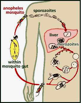
Mosquitoes are known to be annoying. There are more annoying in poverty stricken countries where they spread diseases. The major health issue concerning mosquitoes is the spread of malaria. Researchers at Case Western Reserve University are finding that a blood type in African populations no longer protects against a specific type of malarial infection. Sub-populations in African previously showed a resistance to P. vivax malaria by having a Duffy-negative blood type. Not having the Duffy blood protein disabled the parasite from infecting red blood cells. However, a study was performed on over 600 people from different communities in Madagascar and found that 10% of people exhibiting the disease were, in fact, Duffy-negative.

This new ability of infection in these populations may be due to population mixing. Many people from Southeast Asia now live in Madagascar. These Southeast Asians are Duffy-positive. The children of those from both Duffy-negative and Duffy-positive parents show susceptibility to infections. Peter A. Zimmerman, Ph.D ., a researcher at Case Western Reserve University states that “the study confirms that P. vivax is not dependent on the Duffy antigen for establishing blood-stage infection and disease in Madagascar.” This new finding has a great impact all over Africa, where this natural immunity is the best defense against P. vivax malaria. There are approximately three million new cases of P. vivax malaria infections reported every year.

When mosquitoes bite a person infected with the disease, the malaria causing parasite is taken in as well. The mosquito then bites a healthy person, injecting the parasite into the person’s blood stream. Malaria is one of the “big three” diseases in the world because the parasites are so easily spread. It is particularly endemic in Africa where treatment is limited. There are five types of parasites of parasites that cause malaria. Most anti-malaria campaigns focus on the P. falciparum malarial infection. New efforts must be put in place to fight the P. vivax infections.

(source)

An uncommon use for a common drug offers hope to millions of HIV-positive patients

By Liz H. ‘10

Microscopic view of HIV (green) emerging from an infected T-cell. CDC

A promising new HIV treatment has been discovered in an unlikely source:  a widely available acne medication developed in the 1970s.  A team of scientists from Johns Hopkins University reports that minocyclin stops HIV-infected human cells from reactivating and replicating, in a study published in the April 15th issue of the Journal of Infectious Diseases.  Their findings may lead to an improved and more effective treatment regimen for HIV infection.

The researchers focused their study on latent, non-replicating HIV-infected human T-cells.  T-cells are a type white blood cell that normally fights infection.  HIV infects T-cells and can “rest” inside of them for an extended period of time.  The virus does not harm the T-cell during this latent phase, but can eventually “wake-up” and re-activate the T-cell, which spreads HIV infection and weakens the immune system.

In this study, the scientists treated latent HIV-infected human T-cells with minocycline and measured the level of re-activated T-cells over time.  They also performed the same measurements on cells that were not treated with minocycline.  The researchers found that the minocycline-treated cells did not display detectable levels of reactivation while the untreated cells displayed elevated levels.

Upon closer analysis of the activity of minocycline inside of cells, the scientists discovered that the drug interferes with important cellular communication pathways that cause the T-cell to activate and spread HIV to other cells.  “It prevents the virus from escaping in the one in a million cells in which it lays dormant in a person…That’s the goal:  Sustaining a latent non-infectious state,” explains Gregory Szeto, a Hopkins graduate student who worked on the project.

These findings suggest that minocycline could be used in conjunction with HAART, the current HIV treatment standard, to keep the virus dormant inside of T-cells.  “While HAART is really effective in keeping down active replication, minocycline is another arm of defense against the virus,” says author Janice Clements.  Minocycline is an attractive addition to the current arsenal of HIV medications because it is relatively inexpensive, does not inhibit the ability of T-cells to fight other infections, and is not likely to cause viral drug resistance.

Current treatment for HIV/AIDS involves a combination therapy approach known as HAART.  Patients on HAART take at least 3 antiretroviral drugs daily that act on the virus in different ways to reduce its levels in the bloodstream.  Although HAART can extend the life of an infected individual, it is not a cure and causes unpleasant side effects and the development of drug resistance.  For the 40 million HIV-positive individuals worldwide, this new use for minocycline promises improved outcomes.

Want more information?

What is HIV?

Press Release