Are β-blockers Doing You More Harm Than Good?

Beta-blockers

Chronic Obstructive Pulmonary Disease (COPD) includes a number of diseases where airflow to the lungs is blocked. Cardiovascular diseases are frequent occurrences in patients with COPD.  To manage medical conditions like high blood pressure and abnormal heart rhythm that are associated with cardiovascular diseases, medication called β-blockers are used. However, many physicians shy away from prescribing them due to uncertainty about potential effects of β-blockers on lung activity. This is especially true during episodes of acute COPD exacerbations (sudden worsening of symptoms). Contrary to these conceptions, studies show that β-blockers play a role in alleviating COPD exacerbations and reduce mortality rates.

The TONADO studies conducted by researchers at the Boehringer Ingelheim Pharma GmbH & Co. KG, Ingelheim in Germany conducted research to check lung function in COPD patients receiving bronchodilator (substance that dilate the air passages to the lungs, thus increasing airflow) treatments for a year. FEV1 or forced expiratory volume, a measure of the volume of air that is forced out in 1 second after taking a deep breath, was used to categorize the severity of COPD. Post experimentation, researchers considered data from patients receiving β-blockers to assess the potential effects of these medication on bronchodilator treatment. Researchers assessed these effects throughout the length of the study by analyzing lung function, dyspnea (difficulty breathing) and frequency of exacerbations in COPD patients.

In the study conducted on 5,163 patients, individuals who were on β-blockers showed lower occurrence of COPD with milder symptoms, and higher average FEV1 levels.

Exacerbations were also very low in the  β-blockers group at entry level. One shortcoming of β-blockers was the high occurrence of diseases related to the heart and blood vessels like stroke, heart attack, and cardiac arrhythmia. The improvement in dyspnea seen during bronchodilator treatment and lung function measurements were the same for both groups. Overall, analysis of the obtained data did not show vast negative effects of β-blocker treatment.

The reliability of data from this research is high due to the fact that it was collected over a span of 12 months. In addition, the study had a broad, global population of subjects due to the international nature of the trial. The presence of other additional diseases besides COPD was also taken into consideration during data collection and analysis. Putting all this information together, researchers concluded that their study provides valid information regarding the effects, or lack thereof,  of β-blockers in COPD patients.

In conclusion, the results support β-blocker use. The researchers agreed that the benefits of β-blocker outweigh their potential risks, especially for patients with heart disease, heart failure and hypertension. An analysis of 15 similar studies further supported the findings.

Reference: Maltais, F., et al. 2018. β-Blockers in COPD: A Cohort Study From the TONADO Research Program. Chest.

Link to article

Image: Flickr


Staying Balanced: Sour Taste Buds Linked with the Ventricular System

In January 2018, a study published by the American Association for the Advancement of Science presented a surprising link between the sense of taste and the sense of balance. While trying to determine which genes are responsible for certain taste buds to ascertain sourness, scientists found the same gene at work in the inner ear.

Lemon slices
Lemon slice. Credit: GDJ; Creative Commons Clipart.

When you think “sour,” you might think about puckering at the juice of a slice of lemon, but scientists think about pH levels. Sourness is actually a measure of acidity, due to the fact that a substance is acidic if it contains lots of H+ ions (hydrogen atoms with a positive electrical charge), which is also a mark of low pH. There are different kinds of taste buds: some recognize sweetness, some recognize saltiness, etc. The taste buds that recognize the sour *tang* of Sour Patch Kids contain ion channels that allow H+ ions to flow into the taste bud cell and send a signal to the brain that says, “Wow! This is sour!”

To ascertain which gene or genes are responsible for expressing the proteins necessary for building the H+ ion channels in sour taste bud cells, researchers at the University of Southern California used a mouse model. They compared the transcriptome of mice with sour taste buds with the transcriptome of mice without them. The transcriptome is a collection of all the RNA in a particular cell, and is an indicator of proteins that are being generated and built by a cell. When 41 potential proteins were identified in the sour taste bud cells, but not found in the other taste bud cells, the scientists knew one of them must play a role in the mechanism for detecting sour tastes.

The researchers implanted the potential genes into human embryonic kidney cells (HEK-293) or the female egg cells of a frog model (Xenopus oocytes). Then, the kidney cells and egg cells were flooded with an acidic solution and observed for H+ ion currents. The researchers noticed that the gene Otopetrin1, abbreviated as Otop1, was the only gene to produce an ion channel that permitted H+ ions to pass through.

The gene Otop1 is part of the otopetrin gene family, which happens to be known for the development and function of the vestibular system. The connection is clear when mice with Otop1 mutations exhibited issues with spatial orientation and balance. They could not properly right themselves or swim. Furthermore, the mice with Otop1 mutations had weaker currents of H+ ions in the taste bud cells, which suggests that the mice were not able to fully taste sourness. The scientists at USC hypothesize that Otop1 regulates an optimal pH level in the inner ear during development.

“We never in a million years expected that the molecule that we were looking for in taste cells would also be found in the vestibular system,” senior researcher Emily Liman said. “This highlights the power of basic or fundamental research.”

The Otop1 gene also produces H+ ion channels in the heart, uterus, adrenal gland, mammary gland, and in fat tissue, although the role of H+ ion channels in these regions is not understood. Further research may uncover more intriguing and unanticipated connections within our genetic makeup.

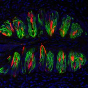
Taste bud cells
Taste bud cells, magnified and artificially colored. The red portions denote cells that detect sour tastes, while the green portions mark cells that detect umami, sweet, or bitter tastes. Credit: Yu-Hsiang Tu and Emily Liman.

Sources:

Tu, Y.H., Cooper, A.J.,Teng, B., Chang, B.R., Artiga, D.J., Turner, H.N., Mulhall, E.M., Ye, W., Smith, A.D., & Liman, E.R. 2018. An evolutionarily conserved gene family encodes proton-selective ion channels. Science [published online] DOI: 10.1126/science.aao3264.

Gersema, E. 2018. Surprising discovery links sour taste to the inner ear’s ability to sense balance. USC Press Room. Retrieved Feb. 18 from http://pressroom.usc.edu/surprising-discovery-links-sour-taste-to-the-inner-ears-ability-to-sense-balance/.

The Beginning of an End to the Autism-Vaccine Debate?

Autism Awareness

Autism spectrum disorder (ASD) is a developmental disorder of the nervous system. The causes of ASD are yet unknown, but it has been linked to both genetic and environmental factors. Researchers at Keele University in the UK have identified aluminum as a potential cause of autism based on a study conducted in 2017 on brain tissue from people diagnosed with ASD.

Aluminum is used in vaccines to enhance the body’s immune response to antigens (harmful or toxic substances). The vaccine-autism debate is highly controversial, but animal models have linked the use of aluminum in vaccines to ASD. The results of this study on human cells further assert these findings.

Aluminum content was measured in 0.3g tissue samples from different regions of the brain of 5 individuals using atomic absorption spectrometry. This method utilizes the difference in the light absorption capabilities of different atoms to find the chemical composition of samples. The values ranged from 1.20 to 4.77μg/g. Past studies have suggested values ≥2.00μg/g as pathologically concerning and those ≥3.00μg/g as pathologically significant. The results showed at least one tissue in each individual that exceeded the established pathologically significant value.

Some of the values recorded were the highest ever measured (17.10, 18.57 and 22.11μg/g).

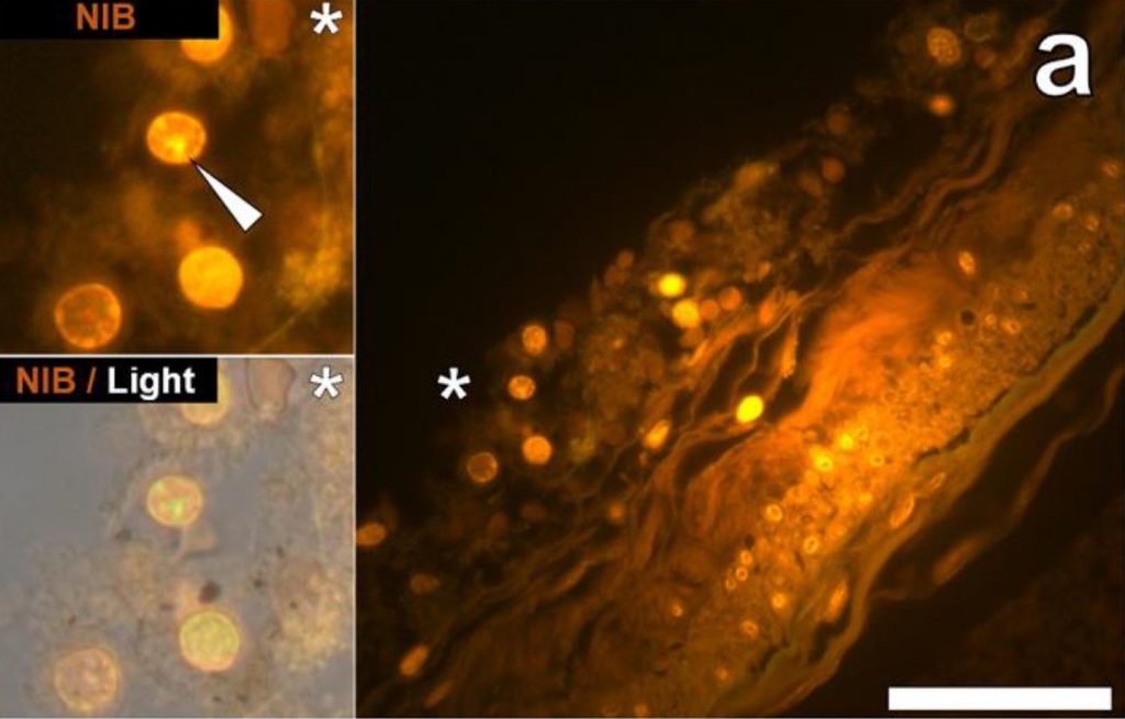
In addition to the concentration, the locations of the aluminum deposits were also examined using fluorescence microscopes. A dye that selectively stains aluminum in cells and human tissues and makes them appear orange or bright yellow was used to view aluminum on the images obtained through the microscope. Deposits were found both inside and outside brain cells. However, the most distinct observation was the presence of metal deposits in the microglia. Microglia are the main immune defense cells inside the central nervous systems and scientists concluded that the deposits seen in them were a direct indication that aluminum had somehow crossed the blood-brain barrier.

fluorescence micrograph
Figure 1 shows the cells in the hippocampus of a 50-year-old male donor used in the study by Mold et al. The white arrow indicated aluminum depositions that were observed via orange fluorescence emission. Hippocampus is the part of the brain considered to be the center of emotion and memory.

Aluminum is toxic to living cells. Although the microglia could remain functional for a certain time period, the metal will eventually show its adverse effects by disrupting this functionality. This directly correlates defective microglia with ASD. In addition to microglia, the study showed aluminum depositions in other tissues from different parts of the brain.

The study also showed great variability in the age groups of donors from 15 to 50 year olds. Initially, the high concentration seen in tissue from a 15 years old donor had greatly puzzled the researchers. However, the evidence of aluminum deposition in the microglia and other intracellular locations ties back to implicate vaccines as a potential cause of ASD and explain how such high amounts of aluminum could have deposited in the brain tissues of a 15 year old boy.

This shows the first ever instance of aluminum concentration measurement in human brain tissues from individuals with ASD. Despite the concrete results, the research was limited due to the lack of a substantial number of subjects and the minimal amount of tissue cells that could be obtained for the study. These factors render the research inadequate by itself to establish ASD as a direct outcome of aluminum deposition from vaccines in brain tissues. However, it is a major stepping stone towards realizing the potential cause of autism spectrum disorder. Now, there is a need for more research to either support or question the results of this study. 

Reference:

Mold, M., Umar, D., King, A., and Exley, C.2018. Aluminium in brain tissue in autism. Journal of Trace Elements in Medicine and Biology 46: 76-82.

Link to article

Link to feature image


Weighing the Risks: Gastric Surgery May Lengthen Life

A recent study found that obese patients who received gastric surgery, rather than nonsurgical weight loss treatment, saw a significant decrease in mortality rates over a 4.5 year period. The researchers identified three specific surgeries, which effectively reduce the size of the stomach so that the patient will feel full with less food. The study consisted of over 8000 obese Israeli citizens and was conducted by the state health service.

The study lasted from 2005-2014, with each patient being followed up with for a minimum of one year after entering the study, an average of 4.5 years and a maximum of 11 years. Specifically, the findings reported a 1.3% mortality rate among obese patients who received gastric surgery and a 2.3% mortality rate among obese patients who opted for nonsurgical treatment. These findings are significant, because even with all the associated risks of surgery, there was still a higher survival rate with it than without.

There is a tendency to dismiss any treatment for obesity other than diet and exercise, as the presence or absence of these are the only treatment or cause of obesity. But in patients who have a history of struggling with this traditional prognosis, they would be better off having the surgery to force them into a lesser diet rather than face the health risks of continuing to remain obese under a less drastic treatment plan, based on these findings.

As a 10 year long study following the ongoing care of 8385 patients, the parameters are more than sufficient to inspire confidence in its results. And even though a 1.3-2.3% may sound small, it is a significant increase in the proportion of mortalities and an indicator of future health and longevity. So perhaps patients who struggle with obesity should consider gastric surgery as a new strategy.


Reges O, Greenland P, Dicker D, Leibowitz M, Hoshen M, Gofer I, Rasmussen-Torvik LJ, Balicer RD. Association of Bariatric Surgery Using Laparoscopic Banding, Roux-en-Y Gastric Bypass, or Laparoscopic Sleeve Gastrectomy vs Usual Care Obesity Management With All-Cause Mortality. JAMA. 2018;319(3):279–290. doi:10.1001/jama.2017.20513

Insight into Pericytes

Blood Brain Barrier and Astrocytes type 1
Blood Brain Barrier. Credit: Ben Brahim Mohammed, Wikimedia Commons

Imagine the vascular system in the brain as a strainer used in cooking. After cooking pasta in a pot of water, you pour the pasta over the strainer, so that it catches the noodles, and the water filters out into the sink. Typically, you want a strainer with small holes, so vegetable pieces or meat pieces cooked with your pasta don’t slip out with the water into the sink.

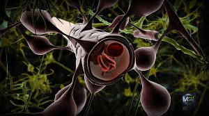
Similarly, specialized cells called pericytes act as the strainer of blood flow in the brain. These cells contribute to forming the blood-brain barrier, which permits nutrients and oxygen to filter through to feed brain cells but prevents toxins from entering the brain. The pericytes play an active role in managing this exchange. Pericytes also regulate blood flow in the small capillary blood vessels. In other words, they determine the width of the blood vessels and decide how much blood can flow freely.

A recent study published in Nature Medicine on February 5th linked pericyte damage with Alzheimer’s Disease and other forms of dementia. Previously, Azheimer’s Disease and other neurodegenerative diseases were associated with accumulations of TAU proteins, toxic proteins that build up over time and inhibit brain function. Researchers at the University of Southern California now think pericytes are to blame as an earlier marker for dementia, causing issues before TAU proteins even show up.

Researchers used a mouse model to simulate pericyte deficiency in humans, and noticed that damaged pericyte cells let some materials leak out of the blood and into the brain that were not supposed to be there, just like a strainer with holes that are too big and macaroni noodles start plopping into the sink. The leaking material was fibrinogen, a protein that creates blood clots at injury sites. During the healing process, fibrinogen is vital, but in the brain, fibrinogen deposits erode away at the insulation barrier of neurons, called myelin, and disrupt electrical communication from one neuron to another. You might equate fibrinogen as the chunks that get through your strainer, and then clog the drain pipe.

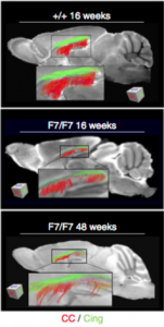
Nerve tracts gradually eroding as the result of damaged pericytes.
Myelin (shown in green and red) gradually erodes away as the result of damaged pericytes.  Credit: Montagne et al.

The alarming discovery was that in the absence of healthy pericytes, fibrinogen leaked into the brain, and the cells that produce myelin, called oligodendrocytes, started to die. By the end of the experiment, 50% of the oligodendrocytes were dying or defective. One hypothesis proposed was that besides directly destroying the oligodendrocytes, fibrinogen also blocks oxygen and nutrients from reaching them, further accelerating cell death.

The scientists are hopeful that their research will initiate new treatments for dementia by focusing on the root of the problem: the damaged pericytes producing leaks in the blood-brain barrier. The senior researcher said, “Perhaps focusing on strengthening the blood-brain barrier integrity may be an answer because you can’t eliminate fibrinogen from blood in humans. This protein is necessary in the blood. It just happens to be toxic to the brain.” With future research, the pericytes may become the primary target for dementia treatment and prevention.

Sources:

Montagne, A., Nikolakopoulou, A., Zhao, Z., Sagare, A.P., Si, G., Lazic, D., Barnes, S.R., Daianu, M., Ramanathan, A., Go, A., Lawson, E.J., Wang, Y., Mack, W.J., Thompson, P.M., Schneider, J.A., Varkey, J., Langen, R., Mullins, E., Jacobs, R.E., & Zlokovic, B.V. 2018. Berichte degeneration causes white matter dysfunction in the mouse central nervous system. Nature Medicine [ePub ahead of print].

Vuong, Zang. 2018. Half of all dementias start with damaged ‘gatekeeper cells.’ USC Press Room. Retrieved Feb. 12 from http://pressroom.usc.edu/half-of-all-dementias-start-with-damaged-gatekeeper-cells/.


Alpha waves, attention, anxiety, oh my!

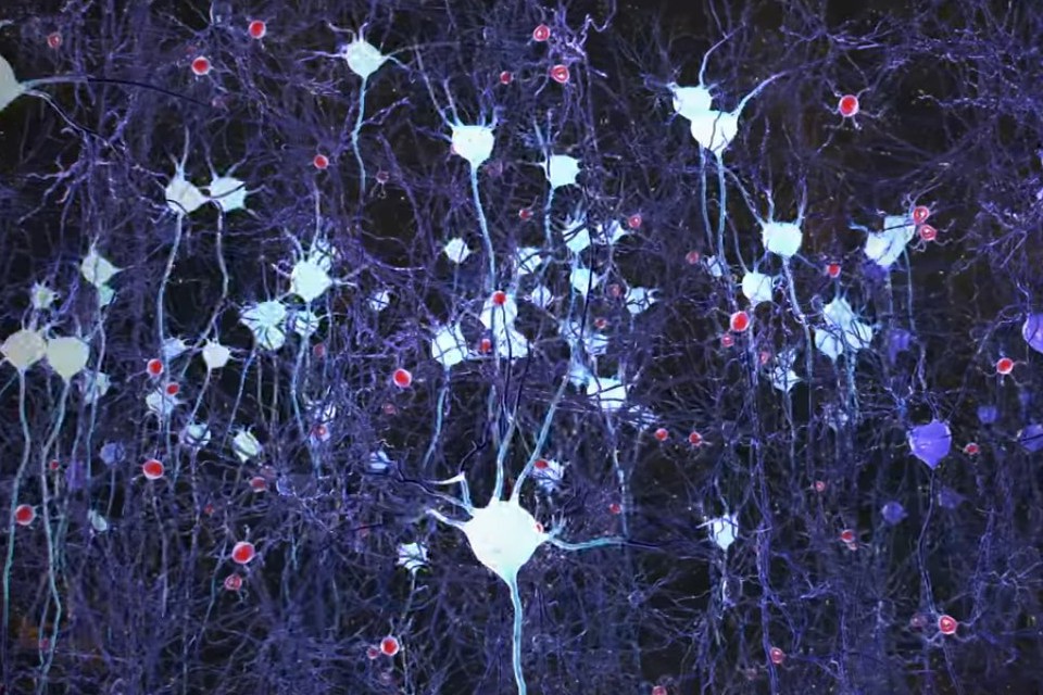
Neurons firing in the brain.
Neurons firing in the brain (artificial color added). Credit: Picower Institute for Learning and Memory, M.I.T.

In a recent study, published in January 2018, scientists pinpointed a unique characteristic of people who experience trait anxiety–differences in alpha brain wave activity. Usually anxiety is correlated with an absence of alpha waves; in anxious brains, beta waves overpower alpha waves, and over time, this accumulates into feelings of constant stress. Researchers in the Departments of Psychology and Psychological Science at Ball State University found that too many alpha waves can create an equally disruptive imbalance.

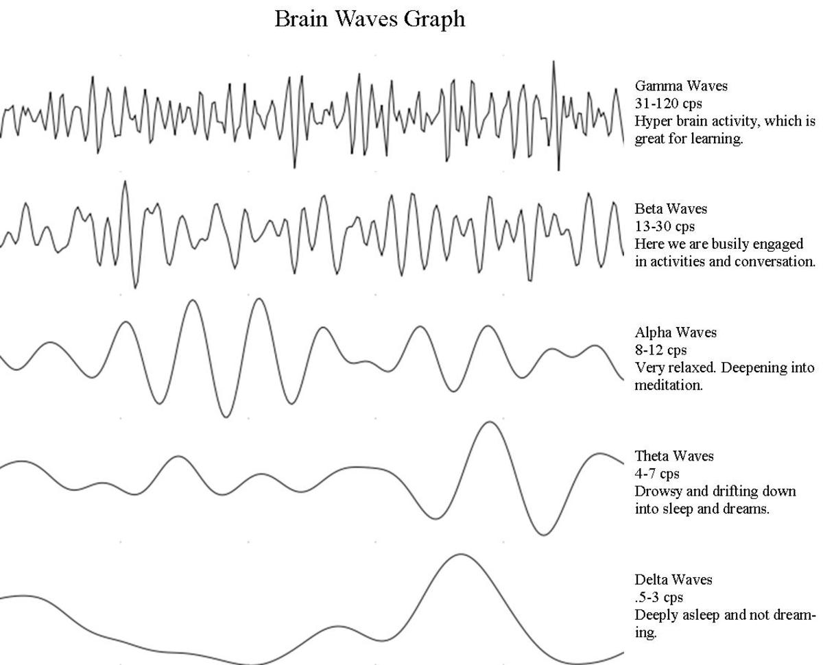
The brain is composed of billions of neurons, which communicate with each other through electrical signaling. When multiple neurons fire simultaneously, they produce electrical oscillations, or “waves.” The frequency of these waves depends on the current level of consciousness: brain waves tend to be lower frequency during deep sleep, but high frequency during problem-solving, decision-making, and other tasks requiring complex thinking and concentration.

Alpha waves, which were evaluated in this study, are known to occur when the mind is in a state of relaxation. At any given moment, the brain might elicit more than one type of brain wave, but alpha waves are most widespread during meditation, while daydreaming, and even during prolonged aerobic activity, like a “runner’s high.” However, as soon as we are alerted with a task, faster beta waves take over.

This may not be the case with highly anxious individuals. Researchers used an EEG to measure the alpha brain waves of a group of individuals in a high-trait anxiety condition, analogous with having an anxiety disorder, and a group of individuals in a low-trait anxiety condition, meaning they showed very few anxiety symptoms. Researchers first measured the alpha waves during a resting, relaxed state, and then while the participants completed a response-inhibition test called the Eriksen-Flanker Task.

Researchers found that the highly anxious individuals demonstrated more alpha wave activity in the resting state, compared to the less anxious individuals. But during the Eriksen-Flanker Task, the two groups demonstrated similar levels of alpha wave activity. In other words, at baseline, the highly anxious individuals were essentially more relaxed than typical, so their brains had to make a further jump to get to an alert and focused state.

While this may seem counter-intuitive, the implications for this experiment are that the prevailing alpha waves in the brain of a highly anxious individual suppress processing of external stimuli and information. The individual might then have trouble focusing on specific tasks and thoughts. In conjunction with previous studies, anxiety has been linked to a lack of alpha waves as well as extra alpha waves in a resting state, suggesting that abnormal alpha brain wave activity alters attention and processing in various ways. More research is needed to more clearly understand this phenomenon, but researchers hope this method of measuring alpha waves will become a tool to measure degrees of anxiety in the future.

brain waves
Types of brain waves, as they appear on an EEG. Credit: Slaven Cvijetic.

Sources:

Ward, R.T., Smith, S.L., Kraus, B.T., Allen, A.V., Moses, M.A., Simon-Dack, S.L. 2018. Alpha band frequency differences between lot-trait and high-trait anxious individuals. NeuroReport 29:79-83.

Bergland, C. 2015. Alpha brain waves boost creativity and reduce depression. Psychology Today. Retrieved Feb 5, 2018 from https://www.psychologytoday.com/blog/the-athletes-way/201504/alpha-brain-waves-boost-creativity-and-reduce-depression


Radiation in Clinical Care

Biomedical Imaging

Medical Intensive care unit (MICU) patients are exposed to radiation higher than annual US Federal occupational standard limits within a short period of time during radioscopic studies. Research conducted between January and December 2013 by scientists at the Cleveland Clinical Foundation discovered this to be true among 3% of the total 4155 patients involved in the study. 

The millisievert (mSv) is a measure of the organic effect of ionizing radiation and is known as the effective dose (ED). Statistical tests conducted on the data obtained in the study showed that 36% of the patients were exposed to radiation higher than the natural background radiation (~3 mSv). Values just 3-5 times higher have been suspected to cause carcinogenesis, although the debate is still ongoing.

Radiation is used in numerous reliable diagnostic procedures in the MICU. Although this has been overshadowing the potential risk of cancers, recently, more medical literatures are exploring its adverse effects. The study also showed an increase in cumulative ED for patients with a higher length of stay at the MICU. Among the radiation based Imaging systems used in healthcare, CT and IR caused the highest amount of radiation burden. However, diagnosis of sepsis, COPD, cirrhosis and Gastrointestinal bleeding were seen to affect cumulative ED. 

radiation burden data distribution
Figure 1 shows the distribution of data obtained by Krishnan et al for radiation burden

Ionizing radiation can lead to cancers by damaging our DNA or RNA, and cause genetic abnormalities. This is especially true for kids and young adults who have a high rate of cellular division and a longer lifespan to express the effects of radiation. Krishnan et al report that despite their adoption of the ALARA (‘as low as reasonably achievable’) principle of radiation safety, patients were exposed to substantial amounts of radiation during diagnosis. Therefore, they suggest methods for assessing the risk vs benefit of radiation therapy.

“Proactive monitoring of CED with real time display in electronic medical resorts will assist physicians in deciding the risk-benefit ratio.”

The study was limited as it was conducted among MICU patients from a single academic medical center. The estimate of ED from previous papers and the limited medical records from which data was obtained could have caused an undervaluation of the cumulative ED. Another shortcoming was the disregard for patient basic characteristics like age and sex. Nevertheless, the results denote a need to conduct more research to weigh the pros and cons of radiation in medicine and its effects on overall patient health.

This makes me wonder, are we so highly dependent on radioscopic resources that we fail to consider the harm it might be doing us? At this day and age of technological advancement, there should be active endeavors to explore the benefits and possible disadvantages of radiation in the medical industry and ways to alleviate them.

Reference: Krishnan S, Moghekar A, Duggal A, Yella J, Narechania S, Ramachandran V, Mehta A, Adhi F, Vijayan AKC, Han X, Dong F, Martin C III, Guzman J, Radiation Exposure in the Medical Intensive Care Unit- Predictors and Characteristics, CHEST (2018), doi: 10. 1016/ j.chest.2018/01/019.

Link to article

Canadian Light Source Research Office


Hormones Do It Again

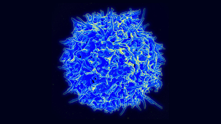
T-cells

Researchers at the UPCM Sorbonne University in France have linked Autoimmune Regulator (AIRE) proteins to the high predisposition of women to autoimmune diseases (AD). Results from numerous studies correlated mutations in AIRE to the occurrence of ADs. Further research was conducted that showed a negative effect of female hormones on the functioning of AIRE.

ADs are results of abnormal immune responses caused by T cells (immune cells in the blood) that recognize body cells as foreign and attack them. AIRE are proteins present in cells of the inner lining of the thymus. They recognize such T cells and eliminate them through apoptosis (cell death) in a natural process called central immune tolerance. To back this hypothesis, research conducted on an autoimmune thyroid mouse model showed an increase in the presence of autoantibodies in the blood upon blocking AIRE expression in the thymus.

T-cells

The relationship between autoantibodies and female sex hormones is most prominent through the high incidence of ADs in females after puberty, a time of significant hormonal differences between males and females.

A study showed that the female sex hormones estrogen and progesterone caused a decrease in the expression of AIRE whereas the hormone DHT (formed from testosterone) showed an increase in expression. Estrogen has been found to contribute to this by causing hypermethylation of the AIRE gene. In this process, methyl groups are added to DNA molecules, hence changing their function without causing significant change in the structure. This is but one method by which female sex hormones lead to ADs. However, there is much more to be learned by exploring the multitude of pathways involved in the relationship between sex hormones, AIRE and ADs.

As an example, in certain body cells, AIRE has been shown to activate genes that are responsible for insulin tolerance by binding to them. Low AIRE expression has been associated with type I diabetes. Besides AIRE, there are other proteins being investigated to check for possible correlations between sex hormones and AD.

PRDM1 (PR domain zinc finger protein 1) is a protein in our body that helps prevent ADs through the deletion of defective T cells in the thymus. Research studies conducted on mice have shown gender dependence on the workings of PRDM1. However, there is no direct evidence of a connection between PRDM1 and autoimmune diseases in humans. There is much more to be explored in regard to these new results.

The reasons behind the gender based difference in AD must surely not be limited to a few proteins or sex hormones. And just how much do our hormones come into play in the determination of our susceptibility to ADs? What factors contribute to strengthen or alleviate this vulnerability? Many such questions can be raised and the answer to them all would be to dig deeper into the mysteries hidden inside of us.

Reference:

Berrih-Aknin, S., Le Panse, R., and Dragin, N.2018. AIRE: a missing link to explain female susceptibility to autoimmune diseases. Annals of the New York Academy of Sciences 1412: 21-32.

Link to article

NASA’s Marshall Space Flight Center

Researchers Stimulate the Amygdala to Stimulate Memory

Think back to your first kiss, your soccer championship game, or hearing about the death of a loved one. Do you remember what you were wearing? Do you remember who was there with you and specifically where you were? You might even remember the exact words from what people said around you. These crystal-clear memories are called flashbulb memories, and are processed by the amygdala, a region of the brain associated with regulating emotions and emotional memory.

A new study now reveals that directly stimulating the amygdala can result in improved memory without a combined emotional experience. Participants in a study at Emory University Hospital received brief, low-amplitude electrical stimulation to the amygdala and demonstrated improved declarative memory the next day without any subjective emotional feelings or involuntary emotional responses, such as increased heart rate or faster breathing.

The study, published online in December 2017 in the journal PNAS, took place in conjunction with the Emory University School of Medicine. Epilepsy patients with electrodes already implanted in their brains were recruited to participate, and fourteen individuals took part in the study. Participants were shown numerous neutral images (i.e. a picture of a basketball or a key), and then either given a short stimulation of the amygdala or no stimulation. Immediately afterwards and again the following day, participants were shown more neutral images in a recognition-memory test. The patients who received the stimulation exhibited greater memory retention of images after one day, compared to the control group.

While the participants in the study were simultaneously receiving treatment for epilepsy, they showed substantial memory enhancement due to the amygdala stimulation. One patient, who suffered from brain damage and memory impairment and rarely recognized researchers and physicians, displayed the most memory improvement. Other patients who experienced seizures in between the initial stimulation and the test the following day showed improved memory, presenting evidence that the amygdala stores memories in spite of other neurologically debilitating disorders. None of the patients reported being able to feel the stimulation.

brain
Illustration of the amygdala (blue), hippocampus (orange), and perirhinal cortex (pink). Credit: Cory Inman, Emory University.

Researchers speculate that the amygdala plays a role in delegating non-emotional declarative memory to other structures, namely the hippocampus and the perirhinal cortex. Specific stimulation to the hippocampus and perirhinal cortex to improve memory has been erratic in prior studies, and the amygdala might be the missing link. According to co-author Joseph Manns, “the long-term goal of this research program is to understand how modulation of the hippocampus by the amygdala can at times lead to memory enhancement and at times lead to memory dysfunction, such as that observed in post-traumatic stress disorder (PTSD).”

Regarding the targeted amygdala stimulation, co-author Cory Inman explained, “One day, this could be incorporated into a device aimed at helping patients with severe memory impairments, like those with traumatic brain injuries or mild cognitive impairment associated with various neurodegenerative diseases.” Small deep-brain stimulation implants are already being used to treat Parkinson’s disease. This study may be utilized in future research to develop similar clinical treatments for patients with memory disorders, so that their non-emotional memories like what they ate for last night’s dinner or what they read in a good book, can be remembered the next day.

Amygdala and hippocampus highlighted in brain
Limbic system imbedded in the brain. Amygdala is shown in red and the hippocampus is shown in purple. Credit: Paul Wissmann, Santa Monica College.

Sources:

Inman, C.S., Manns, J.R., Bijanki, K.R., Bass, D.I., Hamann, S., Drane, D.L., Fasano, R.E., Kovach, C.K., Gross, R.E., and Willie, J.T. 2018. Direct electrical stimulation of the amygdala enhances declarative memory in humans. PNAS 115: 98-103.

Emory Health Sciences. 2017. Direct amygdala stimulation can enhance human memory for a day: Preliminary study of time-specific electrical stimulation. ScienceDaily. Retrieved January 31, 2018 from www.sciencedaily.com/releases/2017/12/171218151808.htm.

Throwing the Baby in With the Bathwater: Underwater Bubble CPAP

ubCPAP

A new method of Continuous Positive Airway Pressure may be less invasive, more effective, and a far lower cost than traditional methods. A CPAP ventilator system is essential for ensuring the normal breathing of newborn, particularly pre-term infants. CPAP has been shown to increase the survival rates of infants and the Underwater Bubble technique would allow it to be used with fewer limitations.


The underwater bubble technique is named such because as air is pumped into the infants lungs, it exhales into a tube the end of which is submerged in a container of water, regulating the pressure of the entire system. Pressure can be adjusted simply by raising or lowering the depth of the tube.


This study did not conduct trials and experiments but rather evaluated the techniques and their effectiveness in a newborn intensive care unit at a Brazilian university hospital.


The underwater bubble method does not require the insertion of a breathing apparatus making it minimally invasive, and is much lower cost than present alternatives, making the technique more readily available to the developing world.


Really the goal of CPAP is to ensure that newborns are able to breathe without the need for mechanical ventilation or intubation, two invasive and high risk techniques that are fortunately becoming less needed as CPAP progresses.


Another study (Dunn 2011) found that CPAP allowed 48% and 54% of newborns to be managed without mechanical respiration or lung inflating fluid.

Sources:

Abelenda V.L.B., Valente T.C.O,. Marinho C.L., and Lopes A.J. 2018 Effects of underwater bubble CPAP on very-low-birthweight preterm newborns in the delivery room and after transport to the neonatal intensive care unit. Journal of Child Health Care: XX(X) 1-12


Dunn MS, Kaempf J, de Klerk A, et al. (2011) Randomized trial comparing 3 approaches to the initial respiratory management of preterm neonates. Pediatrics 128(5): 1069–1076.


http://journals.sagepub.com/doi/pdf/10.1177/1367493517752500

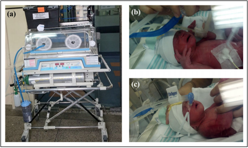
ubCPAP
The Under water Bubble CPAP machine, seen facilitating respiration for a newborn